

Talus Fracture: Treatment, Surgery, and Why These Injuries Are Different
Updated: Aug 27
By Sarang Desai, DO — Fellowship-Trained Orthopedic Foot and Ankle Surgeon
Sports Medicine | McKinney and Flower Mound, Texas
═══════════════════════════════════════
Let me be direct with you: a talus fracture is not like breaking most other bones. The talus has a fragile, easily-injured blood supply, and 57% of its surface is covered in cartilage, so these fractures carry a real risk of two serious complications — avascular necrosis (the bone dying) and post-traumatic arthritis. That's why most displaced talus fractures need surgery, why the timing and technique matter so much, and why I strongly recommend these injuries be handled by a fellowship-trained foot and ankle surgeon.[1][2]
I don't say this to scare you. I say it because I've seen what happens when a talus fracture is underestimated — and I've also seen how well patients can do when it's treated properly from the start. This article explains what makes the talus so unique, how these fractures are treated, and what recovery realistically looks like.
For related reading: The Complete Guide to Ankle Fractures · A Pro Sports Team Physician's Guide to Foot and Ankle Injuries
───────────────────────────────────────
WHAT IS THE TALUS — AND WHY IS IT SO SPECIAL?
The talus is the bone that connects your leg to your foot. It sits between your shinbone (tibia) and your heel bone (calcaneus), and it's the keystone of the ankle. Every step you take, your body weight passes through this one bone.
Three things make the talus fundamentally different from almost every other bone in the body:
- Almost no muscle attachments. Unlike most bones, no muscles attach to the talus. This matters because muscles bring blood supply. Without them, the talus depends on a limited, delicate network of arteries.[2][3]
- It's covered in cartilage. About 57–60% of the talus surface is articular cartilage — the smooth surface that lets joints glide. Cartilage has no blood supply of its own, so there's very little "surface area" left for blood vessels to enter the bone.[1][3]
- A tenuous, retrograde blood supply. The blood supply comes from three arteries (posterior tibial, anterior tibial, and peroneal), and much of it flows "backward" (retrograde) into the talar body through a vulnerable network under the neck. When a fracture disrupts this network, the blood supply to part of the bone can be cut off — and that part can die.[4][3]
That last point is the whole story of why talus fractures are so serious. It's also why the news isn't all bad: research using MRI has shown the blood supply is richer and more redundant than we once thought, with a substantial contribution entering posteriorly — which is exactly why not every talus fracture leads to bone death.[4]
───────────────────────────────────────
HOW DO TALUS FRACTURES HAPPEN?
Talus fractures are rare — less than 1% of all fractures — and they almost always result from high-energy trauma.[5] The classic causes are:
- Car accidents (the foot jammed against the floorboard or pedals)
- Falls from a height (landing on the foot)
- High-impact sports injuries and hard landings
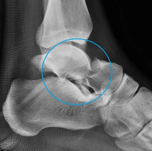
The classic mechanism for a talar neck fracture is the foot being forced violently upward (hyperdorsiflexion), driving the neck of the talus against the tibia.[2]
There's one important exception: the lateral process fracture, often called "snowboarder's ankle." This can happen with a forced dorsiflexion-and-inversion twist and is a well-known injury in snowboarders. It's notorious for being missed on regular X-rays — which is exactly why the right imaging matters so much.[5][6]
───────────────────────────────────────
TYPES OF TALUS FRACTURES
The talus is divided into regions, and where the fracture occurs largely determines the treatment and the risk.
Fracture Location | Key Points | References |
Talar neck | Most common; classified by the Hawkins system, which predicts the risk of bone death (osteonecrosis). Higher grade = higher risk. | |
Talar body | High rate of ankle and subtalar arthritis; crush/comminuted patterns have a poor prognosis. | |
Talar head | Less common; if nondisplaced, can sometimes be treated without surgery. | |
Lateral process ("snowboarder's ankle") | Frequently missed on X-ray; requires a high index of suspicion and CT. | |
Posterior process | Can be subtle; sometimes confused with a normal accessory bone. |
For talar neck fractures, the Hawkins classification is the key tool. It grades the fracture by how displaced and dislocated it is — and the higher the grade, the higher the risk of the bone losing its blood supply and dying.[7][5] Functional outcome worsens as the Hawkins grade increases.[7]
───────────────────────────────────────
HOW TALUS FRACTURES ARE DIAGNOSED
- X-rays are the first step and show most fractures — but they can miss subtle patterns, especially lateral process and posterior process fractures.[5][8]
- CT scan is almost always performed. It's essential for understanding the full extent of the fracture: displacement, comminution (how many pieces), intra-articular extension, and associated injuries. CT is what allows proper surgical planning.[5][2]
- MRI is the most sensitive test for detecting avascular necrosis and is used later if bone death is suspected despite normal X-rays.[9]
If you've had a high-energy foot injury and there's any suspicion of a talus fracture, insist on proper imaging. A missed talus fracture is a genuine problem.
───────────────────────────────────────
NONSURGICAL TREATMENT: WHO QUALIFIES
Here's the reality: only truly nondisplaced (or minimally displaced) fractures can be treated without surgery. For these select cases:[10][5]
- A period of immobilization in a cast or boot, kept non-weight-bearing while the bone heals
- Close follow-up imaging to make sure the fracture doesn't shift
- A gradual, protected return to weight-bearing once healing is confirmed
Even nondisplaced fractures need careful CT evaluation to confirm there's truly no rotation or displacement — because what looks nondisplaced on X-ray sometimes isn't.[5] The vast majority of clinically significant talus fractures are displaced and require surgery.[10]
───────────────────────────────────────
SURGICAL TREATMENT: THE MAINSTAY FOR DISPLACED FRACTURES
For any displaced talus fracture, the goal is anatomic reduction (putting the pieces back in perfect alignment) and stable internal fixation. Getting the alignment exactly right isn't cosmetic — it directly determines how well the ankle and hindfoot function and how likely arthritis becomes.[7][8]
Key principles I follow, consistent with current evidence:
- Reduce dislocations urgently. If the fracture comes with a dislocation, the dislocation should be reduced promptly to take pressure off the skin and remaining blood supply.[7][8]
- The fracture itself doesn't always need to be fixed in the middle of the night. Older teaching was to operate emergently to save the blood supply. Newer evidence suggests that once any dislocation is reduced, delaying definitive fixation until the soft tissues recover can reduce wound complications and infection — and doesn't necessarily change the risk of bone death. What matters most is a perfect, gentle reduction.[8][11]
- Dual-incision approach. For many neck and body fractures, two incisions (anteromedial and anterolateral) allow the surgeon to see both sides and get the alignment perfect. A medial malleolar osteotomy is sometimes added to reach the posterior body.[1]
- Modern fixation. Screws, and increasingly fixed-angle and minifragment plates, provide stable fixation. In select patterns, percutaneous (small-incision) screws work well.[12][1]
- Protect the blood supply. A careful surgical technique that avoids stripping away the remaining blood supply is critical — sloppy dissection can turn a survivable fracture into a dead bone.[11]
This is precisely the kind of surgery where experience and fellowship training matter. The approach, the reduction, the fixation, and the handling of the soft tissues all influence the outcome.
───────────────────────────────────────
THE TWO COMPLICATIONS YOU NEED TO UNDERSTAND
I'm going to be honest about these, because you'll read about them online and I'd rather explain them clearly.
1. Avascular necrosis (osteonecrosis) — the bone dying
This is the complication unique to the talus. When the blood supply is disrupted, part of the talus can die.[13] In a large multicenter study of 798 talus fractures, 42% developed AVN overall — higher after neck fractures (47%) than isolated body fractures (26%).[11] Osteonecrosis has been reported in up to half of talar neck fractures.[8]
But here's the crucial nuance: not every AVN leads to disaster. Many of these bones revascularize (the blood supply grows back) without the talar dome collapsing.[8] There's even a positive radiographic finding — the "Hawkins sign" — that, when present, essentially rules out AVN.[9] The biggest risk factors are the severity of the injury, how displaced it was, open fractures, and — importantly — smoking, which is one of the few risk factors you can control.[11]
2. Post-traumatic arthritis
Because the talus is coated in cartilage and sits in two major joints (the ankle and subtalar joint), damage to those surfaces commonly leads to arthritis over time. Post-traumatic arthritis rates around 43% have been reported, and it's especially common with comminuted body fractures.[14][8]
The takeaway: talus fractures are serious, outcomes depend heavily on injury severity, and a subset of severely injured patients have persistent limitations despite excellent care.[1][14] But an anatomic reduction and careful treatment give you the best possible chance of a good result.
───────────────────────────────────────
RECOVERY: WHAT TO REALISTICALLY EXPECT
Recovery from a talus fracture is a marathon, not a sprint. General expectations:
- Non-weight-bearing period: Typically several weeks to a few months, depending on fracture severity and healing. The talus bears your full body weight, so we don't rush this.
- Gradual weight-bearing: Introduced only once healing is confirmed on imaging.
- Physical therapy: Essential for restoring ankle and subtalar motion, strength, and walking mechanics — similar in principle to rehab after other ankle surgery. Physical Therapy After Ankle Fracture Surgery
- Monitoring for AVN: Follow-up X-rays (and sometimes MRI) over the following months watch for signs of bone death or collapse.[9]
- Full recovery timeline: Often 6–12 months or longer for significant fractures, and some stiffness or activity limitation can persist, particularly after severe injuries.[14]
Return to sport and heavy labor depends heavily on the fracture severity, whether AVN or arthritis develops, and how the ankle and subtalar joints recover. High-demand athletes and workers should plan for a prolonged, staged return.
───────────────────────────────────────
WHAT IF THE TALUS DIES OR ARTHRITIS DEVELOPS?
If AVN progresses to collapse, or if disabling arthritis develops, there are still options — which is one reason not to lose hope:
- Joint-preserving procedures for early disease without collapse (bracing, protected weight-bearing, decompression/revascularization procedures)[13][3]
- Fusion (arthrodesis) of the affected joint to eliminate the painful, damaged surface[13]
- Vascularized bone grafts and advanced salvage techniques that can restore function beyond what was previously possible[1][3]
Treatment for these late complications is highly individualized and best determined case-by-case.[13] This is complex, and it's the kind of problem I manage regularly.
───────────────────────────────────────
COMMON MISTAKES AND MISCONCEPTIONS
- "It's just a sprain." A high-energy foot injury with significant pain and swelling deserves proper imaging. Lateral process fractures in particular masquerade as ankle sprains.[5][6]
- "If X-rays are normal, I'm fine." Not necessarily — X-rays miss subtle talus fractures. CT is often needed.[5]
- "Surgery has to happen tonight or I'll lose the bone." The dislocation needs urgent reduction, but definitive fixation can often be safely delayed until soft tissues recover, which may lower wound complications.[8][11]
- "I can push through and walk on it." The talus carries your body weight; premature weight-bearing risks displacement, malunion, and collapse.
- "Smoking won't affect this." Smoking is a significant, modifiable risk factor for both AVN and nonunion. Stopping genuinely matters.[11]
───────────────────────────────────────
FREQUENTLY ASKED QUESTIONS
1. Why are talus fractures so serious?
Because the talus has a fragile blood supply, no muscle attachments, and is mostly covered in cartilage — so fractures risk avascular necrosis (bone death) and arthritis.[1][3] They also result from high-energy trauma, meaning the injury is severe to begin with.[5]
2. Do all talus fractures need surgery?
No — truly nondisplaced fractures can be treated with immobilization and non-weight-bearing.[10][5] But most clinically significant talus fractures are displaced and require surgery to restore alignment.[7]
3. What is avascular necrosis, and how likely is it?
AVN is death of the bone when its blood supply is cut off. Roughly 42% of talus fractures develop AVN overall, higher with neck fractures and displaced or open injuries — but many revascularize without collapse.[8][11]
4. How long does a talus fracture take to heal?
Significant fractures often take 6–12 months or longer for full recovery, with a prolonged non-weight-bearing period followed by physical therapy.[14]
5. Will I be able to walk normally again?
Many patients do well with anatomic reduction and proper treatment, but outcomes depend on injury severity. A subset of severely injured patients have lasting stiffness or limitation.[1][14]
6. Can a talus fracture be missed?
Yes — especially lateral process ("snowboarder's ankle") and posterior process fractures, which are frequently missed on X-ray and require CT.[5][6]
7. Does the surgery need to happen immediately?
Any dislocation should be reduced urgently. However, definitive fixation can often be delayed until the soft tissues recover, which may reduce wound complications without increasing the risk of bone death.[8][11]
8. What increases my risk of complications?
More severe (higher Hawkins grade) fractures, displacement, open fractures, higher BMI, and smoking. Smoking is the risk factor most within your control.[11]
9. What happens if the bone dies?
Options range from joint-preserving procedures for early disease to fusion, total ankle/talus replacement, or vascularized bone grafts once collapse occurs — individualized to your situation.[13][3]
10. Why should I see a fellowship-trained foot and ankle surgeon?
Talus fractures require precise anatomic reduction, careful protection of the blood supply, correct surgical approach, and long-term monitoring for AVN and arthritis. Experience meaningfully affects outcomes.[1][12]
───────────────────────────────────────
THE BOTTOM LINE
Talus fractures are among the most demanding injuries in foot and ankle surgery. The bone's fragile blood supply and heavy cartilage coating make avascular necrosis and arthritis real risks, and most displaced fractures require surgery to restore precise alignment.[1][7] The good news is that a careful, anatomic reduction — done by a surgeon experienced with these injuries — gives you the best chance at a strong recovery, and even the serious complications have real treatment options today.[1][3]
If you or a loved one has sustained a talus fracture, or you've been told you have one and want an expert opinion, don't wait. Bring your X-rays and CT scan and come in. These are injuries where getting it right early makes all the difference.
───────────────────────────────────────
RELATED READING
───────────────────────────────────────
ABOUT THE AUTHOR
Dr. Sarang Desai is a fellowship-trained orthopedic foot and ankle surgeon with particular expertise in sports medicine. With over 15 years of experience, Dr. Desai serves as a professional sports team physician and treats the full spectrum of foot and ankle conditions — including talus and ankle fractures, cartilage injuries, chronic ankle instability, peroneal tendon disorders, Achilles tendon injuries, Jones fractures, Lisfranc injuries, bunions, foot and ankle arthritis, total ankle replacement, and complex revision surgery.
He is a published researcher, an orthopedic implant inventor, a national lecturer, and a former University of Texas All-American athlete.
Offices in McKinney and Flower Mound, Texas, serving patients throughout Plano, Frisco, Denton, Lewisville, Southlake, Dallas, Fort Worth, Arlington, and the greater Dallas–Fort Worth metroplex and beyond.
SCHEDULE AN APPOINTMENT
📍 McKinney, TX | Flower Mound, TX
If you've sustained a talus fracture and want an expert opinion — bring your X-rays and CT scan and come in. These are injuries where precise, experienced treatment early makes all the difference.
───────────────────────────────────────
This article is for educational purposes only and does not constitute personal medical advice. Always consult a qualified physician about your individual situation.
References
Talus Fractures: Evaluation and Treatment. Lee C, Brodke D, Perdue PW, Patel T. The Journal of the American Academy of Orthopaedic Surgeons. 2020;28(20):e878-e887. doi:10.5435/JAAOS-D-20-00116.
Talus Fractures: All I Need to Know. Al-Jabri T, Muthian S, Wong K, Charalambides C. Injury. 2021;52(11):3192-3199. doi:10.1016/j.injury.2021.10.008.
A chimeric medial femoral condyle chondro‐osseus flap with two thin periosteal flaps to reconstruct partial necrosis of talar body: A case report. Alice Letizia A, Sara T, Stefano B, Mori F, Giulio M. Microsurgery. 2024;44(1):e31127. doi:10.1002/micr.31127.
Quantitative Assessment of the Vascularity of the Talus With Gadolinium-Enhanced Magnetic Resonance Imaging. Miller AN, Prasarn ML, Dyke JP, Helfet DL, Lorich DG. The Journal of Bone and Joint Surgery. American Volume. 2011;93(12):1116-21. doi:10.2106/JBJS.J.00693.
Talar Fractures and Dislocations: A Radiologist's Guide to Timely Diagnosis and Classification. Melenevsky Y, Mackey RA, Abrahams RB, Thomson NB. Radiographics : A Review Publication of the Radiological Society of North America, Inc. 2015 May-Jun;35(3):765-79. doi:10.1148/rg.2015140156.
Hindfoot Fractures: Injury Patterns and Relevant Imaging Findings. Smitaman EE, Davis M. Radiographics : A Review Publication of the Radiological Society of North America, Inc. 2022 May-Jun;42(3):661-682. doi:10.1148/rg.210167.
Surgical Treatment of Talus Fractures. Shakked RJ, Tejwani NC. The Orthopedic Clinics of North America. 2013;44(4):521-8. doi:10.1016/j.ocl.2013.06.007.
Fractures of the Talus: State of the Art. Vallier HA. Journal of Orthopaedic Trauma. 2015;29(9):385-92. doi:10.1097/BOT.0000000000000378.
Avascular Necrosis of the Talus: A Pictorial Essay. Pearce DH, Mongiardi CN, Fornasier VL, Daniels TR. Radiographics : A Review Publication of the Radiological Society of North America, Inc. 2005 Mar-Apr;25(2):399-410. doi:10.1148/rg.252045709.
Adolescent Talus Body Fracture With High Displacement: A Case Report. Hama S, Onishi R, Yasuda M, Minato K, Miyashita M. Medicine. 2018;97(35):e12043. doi:10.1097/MD.0000000000012043.
Identifying Risk Factors for Osteonecrosis After Talar Fracture. Alley MC, Vallier HA, Tornetta P. Journal of Orthopaedic Trauma. 2024;38(1):25-30. doi:10.1097/BOT.0000000000002706.
Talus Fractures: An Update on Current Concepts in Surgical Management. Githens M, Tangtiphaiboontana J, Carlock K, Campbell ST. The Journal of the American Academy of Orthopaedic Surgeons. 2022;30(15):e1015-e1024. doi:10.5435/JAAOS-D-20-01348.
Avascular Necrosis of the Talus. Parekh SG, Kadakia RJ. The Journal of the American Academy of Orthopaedic Surgeons. 2021;29(6):e267-e278. doi:10.5435/JAAOS-D-20-00418.
Conservative and Surgical Treatment of Talar Fractures: A Systematic Review and Meta-Analysis on Clinical Outcomes and Complications. Saravi B, Lang G, Ruff R, et al. International Journal of Environmental Research and Public Health. 2021;18(16):8274. doi:10.3390/ijerph18168274.


Comments