

Pilon Fracture: Distal Tibia Fracture Surgery and Recovery
Updated: Aug 27
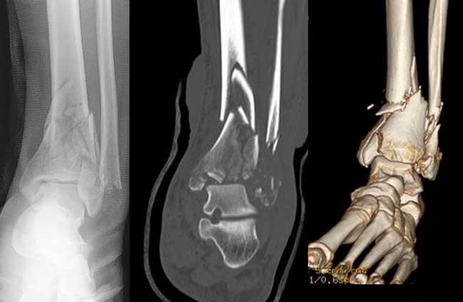
Pilon Fracture: Distal Tibia Fracture Surgery and Recovery
By Sarang Desai, DO — Fellowship-Trained Orthopedic Foot and Ankle Surgeon
Sports Medicine | McKinney and Flower Mound, Texas
═══════════════════════════════════════
I'll be straight with you, because this injury deserves honesty: a pilon fracture is one of the most serious injuries in all of foot and ankle surgery. It's a break of the bottom of the shinbone (the tibial plafond) that shatters into the ankle joint, usually from a high-energy impact, and it comes with severe swelling and soft-tissue damage. Almost all of these need surgery — often done in two stages — and even with excellent, expert care, outcomes can be challenging, with a real risk of stiffness, arthritis, and lasting pain.[1][2] I don't tell you this to discourage you, but so that you understand why the strategy, the timing, and the surgeon's experience matter so enormously with this particular fracture.
Let me explain what's actually happening, why we often operate in two steps, and what recovery genuinely looks like.
For related reading: The Complete Guide to Ankle Fractures · Ankle Fracture-Dislocation: Treatment, Surgery and Recovery
───────────────────────────────────────
WHAT IS A PILON FRACTURE?
"Pilon" is the French word for pestle — the tool that grinds in a mortar. That's exactly what happens here: the talus (the bone of the foot) gets driven up like a pestle into the bottom of the tibia, shattering the weight-bearing joint surface.[3] A pilon fracture (also called a tibial plafond fracture) is a break of the distal tibia that extends into the ankle joint, and it's fundamentally different from a routine ankle fracture — it involves the surface you stand on.[3]
These are uncommon, making up only about 7–10% of tibial fractures, but they punch far above their weight in difficulty.[3] They typically result from:
- Falls from a height (landing on the foot)
- Car crashes (axial load through the foot)
- High-energy sports and industrial injuries
Because the force is so high, pilon fractures usually come with massive swelling, fracture blisters, and sometimes open wounds — and that soft-tissue injury, not just the broken bone, is what drives the entire treatment plan.[1]
───────────────────────────────────────
WHY PILON FRACTURES ARE SO DIFFICULT
There are a few reasons this injury has a reputation among surgeons:
- The skin over the distal tibia is thin and vulnerable. Both the tibia and fibula sit just under the skin at the ankle, with little protective padding. When these fractures swell — and they swell dramatically — operating through that skin too early can lead to catastrophic wound breakdown and infection.[4]
- The joint surface is shattered. In the worst patterns (AO/OTA 43-C3), the joint surface breaks into multiple impacted fragments that are genuinely hard to piece back together anatomically.[5]
- The cartilage is injured at impact. Just like with heel fractures, much of the arthritis risk is set the moment the injury happens, regardless of how good the surgery is.[1]
- Outcomes are humbling. Even with expert care, high rates of post-traumatic arthritis and persistent disability are well documented — this is one of the few fractures where surgeons openly acknowledge the results are often imperfect.[1][6]
───────────────────────────────────────
HOW A PILON FRACTURE IS DIAGNOSED
- X-rays confirm the fracture and show the overall alignment.
- CT scan is essential and is almost always obtained (often after the joint is temporarily stabilized). It maps the individual joint fragments and is what allows a surgeon to plan the precise approach and reduction. In complex fractures, some surgeons even use 3D-printed models of the fracture for planning.[7]
- Soft-tissue assessment is a continuous, hands-on evaluation — the degree of swelling, blistering, and skin condition literally determines when surgery can safely be done.[4]
───────────────────────────────────────
IS SURGERY ALWAYS NEEDED?
For all practical purposes, yes. Historically, nonoperative treatment of displaced pilon fractures led to poor joint reduction, malalignment, and low function.[4] Today, nonsurgical care is reserved only for truly nondisplaced fractures or for patients who can't safely undergo surgery — those with severe medical problems or very low functional demands.[3] The overwhelming majority of pilon fractures are operative injuries.[4]
───────────────────────────────────────
THE STAGED APPROACH: "SPAN, SCAN, PLAN"
This is the single most important concept to understand about modern pilon treatment. For most high-energy pilon fractures, surgeons do not fix the fracture definitively on day one. Instead, they use a staged strategy that trauma surgeons often summarize as "span, scan, plan":[1][4]
Stage 1 — Temporary external fixation (the "span")
Within the first day or so, an ankle-spanning external fixator is applied — a temporary frame with pins in the tibia and foot that holds the leg out to proper length and alignment from the outside, without cutting through the swollen skin. This does two crucial things:[4]
- It restores rough alignment and length, taking pressure off the injured soft tissues.
- It buys time — usually 7 to 21 days — for the swelling to subside and the skin to recover.[4]
During this window, a CT scan is obtained (the "scan") to map the fracture, and the definitive surgery is planned (the "plan").[1]
Stage 2 — Definitive open reduction and internal fixation (ORIF)
Once the soft tissues have recovered — the swelling is down, blisters have healed, and the skin "wrinkles" return — definitive surgery is performed. This is when the joint surface is meticulously rebuilt and held with plates and screws.[4]
Why go to all this trouble? Because the delayed, staged approach was developed specifically to dramatically reduce the wound complications and infections that plagued early attempts to fix these fractures immediately. When surgeons rushed to operate through swollen skin, the results were often disastrous.[5][1]
That said, the timing isn't one-size-fits-all. Acute (immediate) definitive fixation can be safe and effective in the right patient — one with a good soft-tissue envelope, skin wrinkles present, a lower-energy fracture, and no major risk factors like smoking or diabetes.[4] The judgment call between staging and acute fixation is exactly where an experienced surgeon earns their keep.
───────────────────────────────────────
WHAT THE DEFINITIVE SURGERY INVOLVES
The goal is anatomic reduction of the joint surface and restoration of the leg's length and alignment, with the gentlest possible handling of the soft tissues.[3] Key elements:
- Surgical approaches are chosen to match the fracture pattern — most commonly anterolateral, anteromedial, or posterolateral, and often more than one is combined. The incisions are planned carefully around the injured skin.[4]
- The fibula is often fixed first (when broken) to help restore length and act as a scaffold.[3]
- The joint fragments are pieced back together, sometimes with bone graft to support impacted "die-punch" fragments, and held with rafting plates and screws.[5]
- In the worst fractures, some surgeons use modern minimally invasive techniques — such as hexapod ring (fine-wire) fixation with limited internal fixation — which for the most severe C3 patterns has been shown to reduce major complications like deep infection compared with full ORIF.[8]
- In select severe cases — an unsalvageable joint surface, elderly low-demand patients, or poorly controlled diabetes — a primary fusion of the ankle may be the best option from the start.[1]
There's even good evidence for a low-cost safety measure: adding vancomycin powder into the wound at the time of definitive fixation reduces deep gram-positive infections.[1]
Stage | What Happens | Purpose | References |
Emergency | Ankle-spanning external fixator ("span") | Restore length, protect swollen skin | |
Interval (7–21 days) | CT scan, surgical planning; swelling subsides | Map fracture, wait for skin to recover | |
Definitive | ORIF with plates/screws (or ring fixator in severe cases) | Anatomic joint reduction, stable fixation | |
Recovery | Prolonged non/toe-touch weight-bearing + PT | Protect the joint, restore motion |
───────────────────────────────────────
RECOVERY: A LONG, HONEST TIMELINE
I want to set realistic expectations here, because pilon recovery is genuinely one of the longest in foot and ankle surgery.
Typical arc (individualized to your fracture and skin):
- First ~3 weeks: A splint protects the incisions; the ankle is kept elevated to control swelling. Gentle motion exercises often begin once the wound allows.[5]
- Weeks 3–12: This is the defining feature of pilon recovery — a prolonged period of strict toe-touch or non-weight-bearing, commonly lasting about 3 months. The joint surface simply must be protected while it heals.[7][3]
- Around 6 weeks: Partial weight-bearing may begin once early callus (healing bone) appears on X-ray in some protocols.[5]
- Around 3 months: Weight-bearing is progressively advanced as tolerated, under the supervision of physical therapy, once the bone has united.[7][3]
- Physical therapy focuses on range of motion, gait training, strength, and endurance throughout. Physical Therapy After Ankle Fracture Surgery
The bigger picture on recovery is important. A prospective study following pilon patients out to 5 years found that function dropped sharply after injury, improved most between 6 months and 1 year, then continued improving slowly out to 5 years — but function did not fully return to pre-injury baseline, and pain remained a persistent issue. Full recovery is measured in[2] many months to years, not weeks, and longer, dedicated rehabilitation is associated with better outcomes.[6]
───────────────────────────────────────
COMPLICATIONS TO UNDERSTAND
- Post-traumatic arthritis — the most common long-term problem, reported in roughly 16–24% of patients and driven partly by the cartilage damage at impact. Some patients eventually need an ankle fusion or replacement.[1][13]
- Wound problems and infection — the feared early complication in high-energy pilons, which is the entire reason for the staged approach. Deep infection has been reported in around 20% of ORIF-treated severe (C3) fractures in some series.[13][8]
- Nonunion and malunion — the bone failing to heal or healing crooked; nonunion was reported in about 24% of open pilon fractures in one series.[13]
- Stiffness and persistent pain — extremely common and often the most frustrating for patients, as reflected in the long-term outcome data.[2][6]
Modifiable risk factors matter here: smoking, diabetes, and other medical conditions significantly raise the complication risk, and addressing them before surgery genuinely improves outcomes.[1][4]
───────────────────────────────────────
COMMON MISCONCEPTIONS
- "It's just a bad ankle break." No — a pilon fracture breaks the weight-bearing joint surface itself, which makes it far more serious and slower to recover than a typical ankle fracture.[3]
- "They should fix it all right away." For high-energy pilons, rushing definitive surgery through swollen skin is often a mistake; the staged approach exists precisely to avoid wound disasters.[1]
- "I'll be walking in six weeks." Toe-touch or non-weight-bearing for around 3 months is standard. Rushing weight-bearing risks the joint reduction and the healing.[7]
- "Surgery will make my ankle like new." Even with a perfect operation, some arthritis and stiffness are common because of the cartilage injury at impact.[1][2]
- "Smoking won't affect this." It substantially raises the risk of infection and nonunion — stopping is one of the most powerful things a patient can control.[1]
───────────────────────────────────────
FREQUENTLY ASKED QUESTIONS
1. What exactly is a pilon fracture?
It's a high-energy break of the bottom of the shinbone (tibial plafond) that extends into the ankle joint, shattering the weight-bearing surface.[3]
2. How is it different from a regular ankle fracture?
A typical ankle fracture involves the malleoli around the joint; a pilon fracture breaks the joint surface you actually stand on, making it far more serious and prone to arthritis.[3]
3. Does a pilon fracture always need surgery?
Nearly always. Nonsurgical care is reserved for truly nondisplaced fractures or patients who can't safely have surgery.[3][4]
4. Why might I need two surgeries?
High-energy pilons swell dramatically. Surgeons often place a temporary external fixator first to protect the skin, then do the definitive reconstruction 1–3 weeks later once swelling subsides — this dramatically lowers wound complications.[4][1]
5. What is external fixation?
A temporary frame with pins above and below the ankle that holds the leg to length and alignment from the outside, without operating through swollen skin, while you wait for definitive surgery.[4]
6. How long until I can put weight on it?
Typically toe-touch or non-weight-bearing for about 3 months, then progressive weight-bearing as the bone heals — one of the longest protected periods in foot and ankle surgery.[7][3]
7. How long is full recovery?
Many months to years. Function improves most in the first year but continues improving out to 5 years, often without fully returning to pre-injury baseline.[2]
8. Will I get arthritis?
There's a real risk (roughly 16–24%), partly because the cartilage is damaged at the moment of impact. Some patients eventually need a fusion or replacement.[1][13]
9. What raises my risk of complications?
High-energy and open fractures, and modifiable factors like smoking and diabetes. Optimizing these before surgery meaningfully improves outcomes.[1][4]
10. Why see a fellowship-trained foot and ankle surgeon?
Pilon fractures demand precise timing, careful soft-tissue handling, complex reconstruction of the joint surface, and long-term management. Surgeon experience meaningfully affects the outcome of one of the hardest injuries we treat.[1]
───────────────────────────────────────
THE BOTTOM LINE
A pilon fracture is a high-energy break of the ankle joint surface that is serious, slow to recover, and best treated with a carefully staged surgical strategy — often temporary external fixation first to protect the skin, followed by definitive reconstruction once the soft tissues recover.[4][1] Even with expert care, some arthritis, stiffness, and lasting pain are common, and full recovery takes many months to years.[2][6] But a well-timed, anatomic reconstruction by an experienced surgeon gives you the best possible chance at a functional, stable ankle.[3]
If you or a loved one has sustained a pilon fracture, this is an injury where the plan and the surgeon truly matter. Bring your X-rays and CT scan and come in — the earlier the right strategy is in place, the better the ankle does over the long run.
───────────────────────────────────────
RELATED READING
───────────────────────────────────────
ABOUT THE AUTHOR
Dr. Sarang Desai is a fellowship-trained orthopedic foot and ankle surgeon with particular expertise in sports medicine. With over 15 years of experience, Dr. Desai serves as a professional sports team physician and treats the full spectrum of foot and ankle conditions — including pilon fractures, ankle fracture-dislocations, talus and calcaneus fractures, cartilage injuries, chronic ankle instability, peroneal tendon disorders, Achilles tendon injuries, Jones fractures, Lisfranc injuries, bunions, foot and ankle arthritis, total ankle replacement, and complex revision surgery.
He is a published researcher, an orthopedic implant inventor, a national lecturer, and a former University of Texas All-American athlete.
Offices in McKinney and Flower Mound, Texas, serving patients throughout Plano, Frisco, Denton, Lewisville, Southlake, Dallas, Fort Worth, Arlington, and the greater Dallas–Fort Worth metroplex and beyond.
SCHEDULE AN APPOINTMENT
📍 McKinney, TX | Flower Mound, TX
If you've sustained a pilon fracture and want expert care — bring your X-rays and CT scan and come in. With this injury, the right strategy early makes all the difference over the long run.
───────────────────────────────────────
This article is for educational purposes only and does not constitute personal medical advice. Always consult a qualified physician about your individual situation.
References
Management of High-Energy Tibial Pilon Fractures. Murawski CD, Mittwede PN, Wawrose RA, Belayneh R, Tarkin IS. The Journal of Bone and Joint Surgery. American Volume. 2023;105(14):1123-1137. doi:10.2106/JBJS.21.01377.
Long-Term Trajectory of Recovery Following Pilon Fracture Fixation. Middleton SD, Guy P, Roffey DM, et al. Journal of Orthopaedic Trauma. 2022;36(6):e250-e254. doi:10.1097/BOT.0000000000002312.
Medium‐Long‐Term Radiographic and Clinical Outcomes after Surgical Treatment of Intra‐Articular Tibial Pilon Fractures by Three Different Techniques. Biz C, Angelini A, Zamperetti M, et al. BioMed Research International. 2018;2018:6054021. doi:10.1155/2018/6054021.
Left Tibia Pilon Open Fracture Open Reduction and Internal Fixation with External Fixator. Nelson Merchan, MD, Andrew M. Hresko, MD, Edward Kenneth Rodriguez, MD, PhD. Journal of Medical Insight (JOMI).
Treatment of AO/OTA 43‐C3 Pilon Fracture: Be Aware of Posterior Column Malreduction. Guan J, Huang M, Wang Q, Chen Y, Wang L. BioMed Research International. 2019;2019:4265782. doi:10.1155/2019/4265782.
Quality of Life and Functional Impairment After Surgical Treatment of Pilon Fractures-a Case-Control Study With SF-12, EQ-5D-5L and VAS. Gather A, Weigel AC, Bullert B, et al. Journal of Clinical Medicine. 2025;14(19):6965. doi:10.3390/jcm14196965.
The Feasibility of 3D Printing Technology on the Treatment of Pilon Fracture and Its Effect on Doctor‐Patient Communication. Zheng W, Chen C, Zhang C, Tao Z, Cai L. BioMed Research International. 2018;2018:8054698. doi:10.1155/2018/8054698.
Comparison of Complications and Reoperations in AO/OTA 43.C3 Pilon Fractures Treated With Conventional ORIF Versus Minimally Invasive Hexapod Ring Fixation. Bastías GF, Sepúlveda S, Bruna S, et al. Injury. 2023;54 Suppl 6:110884. doi:10.1016/j.injury.2023.110884.
Talus Fractures: Evaluation and Treatment. Lee C, Brodke D, Perdue PW, Patel T. The Journal of the American Academy of Orthopaedic Surgeons. 2020;28(20):e878-e887. doi:10.5435/JAAOS-D-20-00116.
Talus Fractures: All I Need to Know. Al-Jabri T, Muthian S, Wong K, Charalambides C. Injury. 2021;52(11):3192-3199. doi:10.1016/j.injury.2021.10.008.
A chimeric medial femoral condyle chondro‐osseus flap with two thin periosteal flaps to reconstruct partial necrosis of talar body: A case report. Alice Letizia A, Sara T, Stefano B, Mori F, Giulio M. Microsurgery. 2024;44(1):e31127. doi:10.1002/micr.31127.
Quantitative Assessment of the Vascularity of the Talus With Gadolinium-Enhanced Magnetic Resonance Imaging. Miller AN, Prasarn ML, Dyke JP, Helfet DL, Lorich DG. The Journal of Bone and Joint Surgery. American Volume. 2011;93(12):1116-21. doi:10.2106/JBJS.J.00693.
Open Versus Closed Pilon Fractures: Comparison of Management, Outcomes, and Complications. Lu V, Zhang J, Zhou A, et al. Injury. 2022;53(6):2259-2267. doi:10.1016/j.injury.2022.03.018.


Comments