

Calcaneus Fracture: When Does a Broken Heel Need Surgery?
Updated: Aug 27
═══════════════════════════════════════
Calcaneus Fracture: When Does a Broken Heel Need Surgery?
By Sarang Desai, DO — Fellowship-Trained Orthopedic Foot and Ankle Surgeon
Sports Medicine | McKinney and Flower Mound, Texas
═══════════════════════════════════════
Here's the honest answer up front: not every broken heel needs surgery, and the decision is genuinely one of the most nuanced in all of foot and ankle surgery. Undisplaced fractures are almost always treated without surgery.[1] For displaced fractures that break into the joint — the serious ones — the evidence is legitimately mixed: surgery can restore the heel's shape and may offer a small functional benefit, but it carries a real risk of wound and infection complications, and a landmark trial found no clear advantage over nonsurgical care at two years for typical fractures.[2][1] That's exactly why this decision should be made carefully, with a surgeon who does these regularly.[3]
I want to walk you through this the way I would in clinic, because "should I have surgery on my heel?" deserves a real explanation — not a one-size-fits-all answer.
For related reading: The Complete Guide to Ankle Fractures · Talus Fracture: Why These Injuries Are Different
───────────────────────────────────────
WHAT IS A CALCANEUS FRACTURE?
The calcaneus is your heel bone — the largest bone in the foot and the foundation your body weight lands on with every step. A broken heel typically happens from a fall from a height or a high-impact event like a car crash, and it's more common in young, working-age adults.[1] It's a painful, disabling injury that often keeps people from bearing weight for many weeks.[1]
The critical distinction is whether the fracture enters the subtalar joint (the joint between the heel bone and the talus above it):
- Extra-articular fractures — the break spares the joint surface. These generally do well without surgery.[1]
- Intra-articular fractures — the break extends into the subtalar joint. These are the difficult ones, and they make up the majority of significant calcaneus fractures. When the joint surface is displaced, we call it a displaced intra-articular calcaneal fracture (DIACF).[1]
───────────────────────────────────────
WHY THESE FRACTURES ARE SO CHALLENGING
A few things make calcaneus fractures notoriously difficult:
- The soft-tissue envelope is fragile. The skin over the heel has a precarious blood supply. Massive swelling, blisters, and fracture blisters are common, and operating through compromised skin risks catastrophic wound breakdown and infection. The condition of the soft tissues drives both whether and when we operate.[4][3]
- The cartilage is often damaged at the moment of impact. Studies of the cartilage from these fractures show chondrocyte (cartilage cell) viability drops significantly after the injury — meaning some arthritis is "baked in" from the trauma itself, no matter how perfect the surgery.[5]
- Restoring the heel's 3D shape is hard. Malunion — a heel that heals wide, short, and misaligned — causes shoe-fitting problems, tendon impingement on the outside of the ankle, and altered mechanics. Restoring the heel's height, width, and joint congruity is the whole goal of surgery.[1][6]
───────────────────────────────────────
HOW CALCANEUS FRACTURES ARE DIAGNOSED
- X-rays are the starting point and let us measure Böhler's angle — a key indicator of how collapsed the heel is.[7]
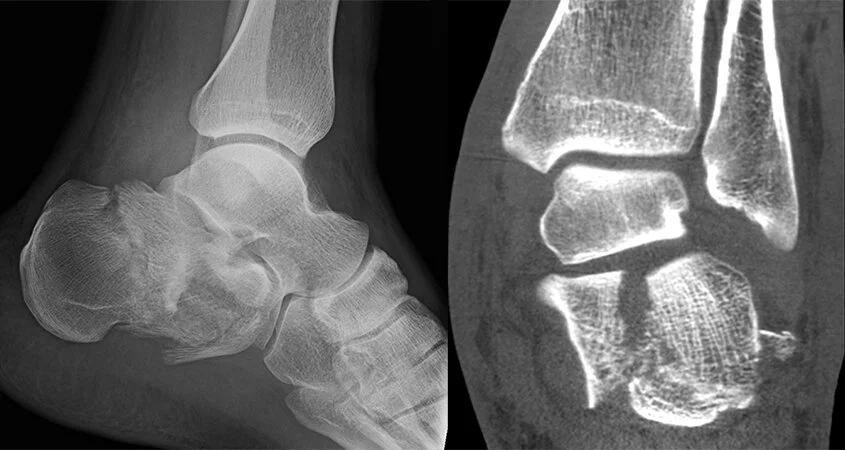
- CT scan is essential. Plain X-rays can't fully characterize these fractures. CT reveals the fracture pattern, joint involvement, and comminution, and it's used for the Sanders classification (based on the number of joint fragments), which guides treatment and predicts outcome.[3]
Higher Sanders grades (more joint fragments) generally mean worse outcomes and factor heavily into decision-making.[8]
───────────────────────────────────────
NONSURGICAL TREATMENT: WHO QUALIFIES
Nonsurgical management is the clear choice for:
- Undisplaced extra-articular fractures — managed with non-weight-bearing for about 4–6 weeks, rest, ice, compression, and elevation, then gradual mobilization.[1]
- Many displaced intra-articular fractures, particularly in patients where the risks of surgery outweigh the benefits.
Nonsurgical care means rest, elevation, ice for swelling, sometimes a cast or removable splint, and a period of not putting weight on the leg — typically at least six weeks.[1] The historical downside is that a displaced fracture left alone can heal malunited — wide, stiff, and painful — sometimes requiring later reconstruction.[1]
───────────────────────────────────────
WHAT THE EVIDENCE ACTUALLY SHOWS ABOUT SURGERY
This is where I want to be straight with you, because the science matters.
The UK Heel Fracture Trial — a rigorous multicenter randomized trial of 151 patients — found no significant difference in pain or function at two years between surgery (ORIF) and nonsurgical treatment for typical displaced intra-articular fractures, and complications and reoperations were significantly more common in the surgical group (odds ratio 7.5).[2] Based on those findings, the authors did not recommend routine ORIF for these fractures.[2]
However, the picture isn't one-sided:
- The 2023 Cochrane review concluded that the evidence — albeit low certainty — would support considering surgery for at least a small benefit in patient-reported outcomes, at the cost of increased complication risk.[1]
- Other studies show that when an anatomic reduction is achieved and Böhler's angle is restored, outcomes are good, and better than nonsurgical care in those specific patients.[7]
- The likelihood of a good-to-excellent result rises when anatomic reduction is obtained.[3]
The reconciliation: surgery helps most when it reliably restores the anatomy — and that depends on the fracture pattern, the soft tissues, the patient, and the surgeon's experience.[3]
───────────────────────────────────────
SO — WHEN DOES A BROKEN HEEL ACTUALLY NEED SURGERY?
Putting the evidence together, surgery is most strongly considered for:
- Displaced intra-articular fractures in younger, active, healthy patients where restoring joint congruity and heel shape offers the best long-term function[7][3]
- Significant loss of heel height/Böhler's angle or a widened heel that would cause malunion, shoe problems, and tendon impingement if left alone[1]
- Fracture-dislocations and severely displaced tongue-type fractures where the skin is at risk from bone pressure — these can be surgical urgencies to prevent skin death[3]
- Open fractures, which require prompt surgical debridement[3]
- Some Sanders IV (highly comminuted) fractures, where primary subtalar fusion is sometimes considered — though this should be used cautiously given high revision rates in some series[8]
Surgery is often best AVOIDED or approached very cautiously in:
- Smokers — non-union and wound complications are markedly higher; in one series non-union occurred only in smokers[8]
- Diabetics, patients with peripheral vascular disease, or a badly compromised soft-tissue envelope[4][3]
- Undisplaced fractures[1]
A critical principle across all of this: timing follows the soft tissues. Surgery is typically delayed until swelling subsides and skin wrinkles return — often 1–2 weeks — to minimize wound complications.[1][3]
───────────────────────────────────────
THE SURGERY ITSELF — AND A MAJOR ADVANCE
The goal of surgery is to restore the heel's height, width, alignment, and joint surface congruity, then hold it with plates and screws.[6]
The big shift in recent years is the surgical approach:
- Extensile lateral approach — the traditional large L-shaped incision. It gives excellent exposure but carries the highest risk of wound breakdown and infection.[3]
- Sinus tarsi and percutaneous (minimally invasive) approaches — smaller incisions that have made prompter surgery with lower complication rates possible. This is a genuine advance and is increasingly how these are done.[6]
- Primary subtalar fusion — reserved for the most severe (Sanders IV) fractures with unreconstructable joints, and used with caution.[1][8]
The move toward minimally invasive techniques is exactly why the older "surgery isn't worth the risk" data may not fully apply to modern, well-selected cases.[1][6]
───────────────────────────────────────
RECOVERY: WHAT TO REALISTICALLY EXPECT
Recovery from a heel fracture — surgical or not — is a long road:
- Non-weight-bearing: Traditionally 6–10 weeks for both surgical and nonsurgical patients. Importantly, newer evidence suggests earlier weight-bearing (starting protected weight-bearing before six weeks) does not worsen outcomes and is increasingly favored after stable fixation.[1][9][10]
- Range-of-motion exercises: Started early after surgery in many protocols to preserve subtalar and ankle motion.[10]
- Physical therapy: Essential for restoring motion, strength, and gait. Similar in principle to rehab after other hindfoot surgery. Physical Therapy After Ankle Fracture Surgery
- Full recovery: Often 6–12 months or longer, and some stiffness, swelling, and activity limitation can persist — particularly after severe fractures.[11][3]
Return to physically demanding work and sports is often the slowest milestone, and some patients with severe injuries don't return to their pre-injury activity level.[11]
───────────────────────────────────────
COMPLICATIONS YOU SHOULD UNDERSTAND
- Post-traumatic subtalar arthritis — the most common long-term problem, driven partly by the cartilage damage from the initial impact. Rates as high as 77% have been reported in surgical series, with roughly 18% eventually needing a subtalar fusion. Successful salvage with subtalar arthrodesis is well established.[8][5][3]
- Wound-healing problems and infection — the most feared early complications, especially with the extensile approach and in smokers/diabetics.[8][4]
- Malunion — a wide, short, misaligned heel causing shoe problems and tendon impingement.[1]
- Nerve irritation (sural nerve), heel pad pain, and stiffness.[4]
- Non-union — rare, and in one series occurred only in smokers.[8]
───────────────────────────────────────
COMMON MISCONCEPTIONS
- "A broken heel always needs surgery." No — undisplaced fractures heal without surgery, and even many displaced ones do reasonably well nonsurgically.[1][2]
- "Surgery will make it good as new." Some arthritis is determined by the cartilage damage at the moment of impact, regardless of a perfect operation.[5]
- "They should operate right away." Except for open fractures or skin at risk, surgery is usually delayed until swelling settles to protect the skin.[1][3]
- "Smoking won't matter." It matters a great deal — smoking substantially raises the risk of wound problems and non-union.[8]
- "I'll be walking in a couple weeks." Heel fractures typically require weeks of protected weight-bearing and months to fully recover.[1][11]
───────────────────────────────────────
FREQUENTLY ASKED QUESTIONS
1. Does every broken heel need surgery?
No. Undisplaced fractures are treated without surgery, and even displaced intra-articular fractures don't always benefit from it — a major trial found no clear advantage over nonsurgical care at two years for typical fractures.[1][2] The decision is individualized.
2. When is surgery clearly recommended?
For open fractures, fracture-dislocations with skin at risk, and displaced intra-articular fractures in younger, active, healthy patients where restoring anatomy offers the best long-term function — especially when significant heel collapse or widening is present.[7][3]
3. Who should probably avoid surgery?
Smokers, diabetics, patients with poor circulation or a badly swollen/compromised soft-tissue envelope, and those with undisplaced fractures.[8][4][3]
4. Why isn't surgery done immediately?
Because operating through swollen, blistered skin risks serious wound breakdown. Surgery is usually delayed 1–2 weeks until swelling subsides — unless the fracture is open or the skin is at risk.[1][3]
5. What does the surgery involve?
Restoring the heel's height, width, and joint surface, then fixing it with plates and screws — increasingly through minimally invasive sinus tarsi or percutaneous approaches that lower complication rates.[6]
6. How long until I can walk on it?
Traditionally 6–10 weeks of non-weight-bearing, though newer evidence supports earlier protected weight-bearing after stable fixation without worsening outcomes.[1][9][10]
7. How long is full recovery?
Often 6–12 months or longer, with some stiffness or limitation possible after severe injuries.[11][3]
8. Will I get arthritis?
Post-traumatic subtalar arthritis is common — reported as high as 77% in some surgical series — partly because cartilage is damaged at impact. If it becomes disabling, a subtalar fusion is a reliable salvage.[8][5][3]
9. What's the biggest risk factor I can control?
Smoking. It dramatically increases wound complications and non-union — stopping genuinely improves your odds.[8]
10. Why see a fellowship-trained foot and ankle surgeon?
Outcomes depend heavily on accurate reduction, soft-tissue handling, and surgeon/institutional experience. This is a decision — and an operation — where expertise meaningfully affects results.[3]
───────────────────────────────────────
THE BOTTOM LINE
Whether a broken heel needs surgery depends on the fracture, the soft tissues, and the patient. Undisplaced fractures heal without surgery. Displaced intra-articular fractures require a careful, individualized decision — the evidence shows surgery can restore anatomy and offer a small functional benefit but carries real complication risks, and it helps most when it reliably restores the heel's shape in the right patient.[2][1][3] Modern minimally invasive techniques have improved the risk-benefit balance, and smoking cessation and proper timing dramatically improve outcomes.[6][8]
If you've broken your heel, or you've been given conflicting advice about surgery, this is exactly the kind of decision worth an expert opinion. Bring your X-rays and CT scan and come in — the right plan for your heel depends on the details.
───────────────────────────────────────
RELATED READING
───────────────────────────────────────
ABOUT THE AUTHOR
Dr. Sarang Desai is a fellowship-trained orthopedic foot and ankle surgeon with particular expertise in sports medicine. With over 15 years of experience, Dr. Desai serves as a professional sports team physician and treats the full spectrum of foot and ankle conditions — including calcaneus, talus, and ankle fractures, cartilage injuries, chronic ankle instability, peroneal tendon disorders, Achilles tendon injuries, Jones fractures, Lisfranc injuries, bunions, foot and ankle arthritis, total ankle replacement, and complex revision surgery.
He is a published researcher, an orthopedic implant inventor, a national lecturer, and a former University of Texas All-American athlete.
Offices in McKinney and Flower Mound, Texas, serving patients throughout Plano, Frisco, Denton, Lewisville, Southlake, Dallas, Fort Worth, Arlington, and the greater Dallas–Fort Worth metroplex and beyond.
SCHEDULE AN APPOINTMENT
📍 McKinney, TX | Flower Mound, TX
If you've broken your heel and want an expert opinion on whether surgery is right for you — bring your X-rays and CT scan and come in. This is a decision where the details, and experience, truly matter.
───────────────────────────────────────
This article is for educational purposes only and does not constitute personal medical advice. Always consult a qualified physician about your individual situation.
References
Surgical Versus Non-Surgical Interventions for Displaced Intra-Articular Calcaneal Fractures. Lewis SR, Pritchard MW, Solomon JL, Griffin XL, Bruce J. The Cochrane Database of Systematic Reviews. 2023;11:CD008628. doi:10.1002/14651858.CD008628.pub3.
Operative Versus Non-Operative Treatment for Closed, Displaced, Intra-Articular Fractures of the Calcaneus: Randomised Controlled Trial. Griffin D, Parsons N, Shaw E, et al. BMJ (Clinical Research Ed.). 2014;349:g4483. doi:10.1136/bmj.g4483.
Current Concepts Review: Intra-Articular Fractures of the Calcaneus. Epstein N, Chandran S, Chou L. Foot & Ankle International. 2012;33(1):79-86. doi:10.3113/FAI.2012.0079.
Complications of Intraarticular Calcaneal Fractures. Lim EV, Leung JP. Clinical Orthopaedics and Related Research. 2001;(391):7-16. doi:10.1097/00003086-200110000-00003.
Chondrocyte Viability After Intra-Articular Calcaneal Fractures in Humans. Ball ST, Jadin K, Allen RT, et al. Foot & Ankle International. 2007;28(6):665-8. doi:10.3113/FAI.2007.0665.
Calcaneus Fractures: A Review of Management, Treatment, and Recent Advances. Schneiderman BA, Carlson BA, Johnson JP, Elsissy JG. The Journal of the American Academy of Orthopaedic Surgeons. 2026;34(6):e821-e831. doi:10.5435/JAAOS-D-24-00567.
A Comparative Study of Operative and Conservative Treatment of Intraarticular Displaced Calcaneal Fractures. Kamath KR, Mallya S, Hegde A. Scientific Reports. 2021;11(1):3946. doi:10.1038/s41598-021-83636-9.
Outcomes of Intra-Articular Calcaneal Fractures: Surgical Treatment of 114 Consecutive Cases at a Maximum Care Trauma Center. Schindler C, Schirm A, Zdravkovic V, et al. BMC Musculoskeletal Disorders. 2021;22(1):234. doi:10.1186/s12891-021-04088-w.
The Effect of Time to Post-Operative Weightbearing on Functional and Clinical Outcomes in Adults With a Displaced Intra-Articular Calcaneal Fracture; A Systematic Review and Pooled Analysis. De Boer AS, Van Lieshout EMM, Van Moolenbroek G, Den Hartog D, Verhofstad MHJ. Injury. 2018;49(4):743-752. doi:10.1016/j.injury.2018.02.021.
Calcaneal Fracture: Results of Earlier Rehabilitation After Open Reduction and Internal Fixation. Park ES, Choi Y, Lee J, Park SH, Lee HS. Archives of Orthopaedic and Trauma Surgery. 2021;141(6):929-936. doi:10.1007/s00402-020-03575-4.
Open Hindfoot Injuries. Lawrence SJ, Singhal M. The Journal of the American Academy of Orthopaedic Surgeons. 2007;15(6):367-76. doi:10.5435/00124635-200706000-00006.


Comments