

Ankle Cartilage Injury (Osteochondral Lesion of the Talus): A Foot and Ankle Surgeon's Complete Guide
Updated: Aug 27
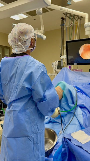
You sprained your ankle. You did the right things — rest, ice, maybe even physical therapy. The swelling went down. The bruising faded. But months later, something still isn't right.
There's a deep ache inside the ankle joint. It's worse after activity. Sometimes the ankle catches or locks. Sometimes it swells after a long run or a hard practice. You've been told it's "just a sprain" — but sprains don't usually hurt like this for this long.
If this sounds like you, there's a good chance you have an osteochondral lesion of the talus — a cartilage injury inside the ankle joint. And you're far from alone. Up to 50% of acute ankle sprains and fractures result in some form of cartilage damage to the talus, and many of these injuries go undiagnosed for months or even years.
I'm Dr. Sarang Desai, a fellowship-trained orthopedic foot and ankle surgeon in McKinney and Flower Mound, Texas. As a professional sports team physician and published researcher, I treat ankle cartilage injuries in everyone from professional athletes to weekend runners to people who just want to walk without pain. This is one of the most complex problems in foot and ankle surgery — and one of the most rewarding to treat when it's done right.
This guide will explain what's actually happening inside your ankle, why it matters, how we diagnose it, and what the treatment options are — from conservative management to the most advanced cartilage restoration techniques available today.
Related guides:
---
What Is an Osteochondral Lesion of the Talus?
Let me break this term down because it sounds intimidating but the concept is straightforward.
Osteo = bone. Chondral = cartilage. Lesion = injury. Talus = the bone that sits inside the ankle joint.
So an osteochondral lesion of the talus (OLT) is an injury to the cartilage surface and the underlying bone of the talus — the dome-shaped bone that your tibia (shinbone) sits on top of. You may also hear this called an osteochondral defect (OCD), osteochondritis dissecans, a talar dome lesion, or simply an ankle cartilage injury. They all refer to the same basic problem.
The cartilage on the talar dome is only about 1-2 millimeters thick, but it's critically important. It provides a smooth, nearly frictionless surface for the ankle joint to glide on. When this cartilage is damaged — whether from a single traumatic event or from repetitive microtrauma — the result is pain, swelling, catching, and progressive joint damage.
Here's what makes cartilage injuries so challenging: cartilage has almost no blood supply. Unlike bone, which heals reliably because blood brings healing cells and nutrients to the injury site, cartilage has very limited ability to repair itself. A cartilage injury that's left untreated doesn't get better on its own in most cases — it either stays the same or gets worse.
---
Why Did This Happen?
Trauma (The Most Common Cause)
The majority of talar osteochondral lesions are caused by ankle trauma — specifically ankle sprains and ankle fractures. When the ankle rolls, the talus impacts against the tibia or fibula, and the cartilage surface gets crushed, cracked, or sheared off.
Osteochondral lesions occur in up to 70% of ankle sprains and fractures. Many of these are small and asymptomatic — the patient recovers from the sprain and never knows the cartilage was damaged. But in a significant number of cases, the cartilage injury becomes the primary source of ongoing pain.
This is why I always tell patients: if your ankle sprain pain hasn't resolved after 3-4 months of appropriate treatment, it's not "just a sprain" anymore. Something else is going on — and a cartilage lesion is at the top of the list.
Chronic Ankle Instability
This is the second most common cause, and it's closely related to trauma. When the ankle ligaments are torn and don't heal properly, the ankle becomes unstable. Every time it gives way, the talus shifts abnormally inside the joint, creating repetitive microtrauma to the cartilage surface.
As I discuss in my chronic ankle instability guide, one in three patients with chronic ankle instability has a cartilage lesion inside the joint. This is one of the main reasons I take ankle instability seriously — it's not just about the inconvenience of a wobbly ankle, it's about protecting the cartilage.
Atraumatic Causes
In some patients, osteochondral lesions develop without any clear history of trauma. These may be related to:
- Vascular insufficiency — reduced blood supply to a portion of the talar dome
- Genetic predisposition — some patients seem prone to cartilage breakdown
- Repetitive stress — high-impact activities over time
- Malalignment — abnormal foot mechanics that concentrate stress on one area of the talus
Atraumatic lesions tend to be on the medial (inside) aspect of the talus and are often deeper, with more subchondral bone involvement (cysts).
Where Do These Lesions Occur?
Medial (inside) lesions account for about 67% of all talar osteochondral lesions. They tend to be deeper, more cup-shaped, and are often associated with subchondral cysts. They're frequently atraumatic or related to chronic instability.
Lateral (outside) lesions account for about 33%. They tend to be shallower, more wafer-shaped, and are more commonly associated with acute trauma (a specific ankle sprain or fracture).
The location matters because it affects surgical approach, treatment options, and prognosis.
---
Symptoms: What Does an Ankle Cartilage Injury Feel Like?
The symptoms of an osteochondral lesion can be frustratingly vague, which is one reason these injuries are often missed or misdiagnosed. Here's what patients typically describe:
Deep ankle pain. Not the sharp, superficial pain of a ligament sprain on the outside of the ankle. This is a deep, aching pain that feels like it's coming from inside the joint. Patients often point to the front of the ankle or can't pinpoint exactly where it hurts.
Activity-related pain. The pain is typically worse with weight-bearing activities — walking, running, jumping, going up and down stairs. It often improves with rest but comes back as soon as you're active again.
Swelling. Intermittent swelling inside the ankle joint, especially after activity. This is different from the lateral (outside) swelling you see with a ligament sprain.
Catching, clicking, or locking. If a piece of cartilage or bone has partially or completely separated from the talar dome, it can act as a loose body inside the joint, causing mechanical symptoms — the ankle catches, clicks, or briefly locks.
Stiffness. The ankle feels stiff, especially in the morning or after sitting for a long time.
Giving way. Some patients describe a sensation of the ankle giving way — but this is different from the lateral instability of a ligament injury. It's more of a "the ankle doesn't feel right" sensation.
The key pattern: Pain that persists months after an ankle sprain, is deep inside the joint, worsens with activity, and is associated with intermittent swelling or catching — this is a cartilage lesion until proven otherwise.
---
How Is an Ankle Cartilage Injury Diagnosed?
Physical Exam
The physical exam for an osteochondral lesion is often less dramatic than for other ankle injuries. There may be:
- Mild joint effusion (swelling inside the joint)
- Tenderness along the anterior joint line (front of the ankle)
- Pain with forced dorsiflexion and plantarflexion (pushing the ankle up and down)
- Pain with axial loading (pushing straight up through the ankle)
- A positive anterior drawer test if there's associated ligament instability
I also evaluate for associated conditions that frequently coexist — chronic ankle instability, peroneal tendon disorders, and hindfoot malalignment.
Imaging
X-rays. Weight-bearing ankle X-rays are the starting point. They can show larger osteochondral lesions, loose bodies, and associated arthritis. However, many cartilage lesions — especially smaller ones — are invisible on X-rays.
MRI. This is the most important imaging study for diagnosing osteochondral lesions. MRI shows:
- The size and location of the cartilage defect
- The depth of bone involvement
- The presence of subchondral cysts
- Bone marrow edema (a sign of active injury)
- The condition of the overlying cartilage (intact, fissured, or detached)
- Associated pathology (ligament tears, tendon injuries, loose bodies)
MRI is essential for treatment planning because the size, depth, and location of the lesion determine which surgical technique is most appropriate.
CT scan. CT provides excellent detail of the bony architecture and is particularly useful for:
- Measuring the exact dimensions of the lesion (length, width, depth)
- Evaluating subchondral cysts
- Surgical planning for osteochondral transplantation
- CT arthrography (CT with contrast injected into the joint) is considered the gold standard in some centers for classifying lesion morphology
CT arthrography enables morphologic analysis and classification of the lesion into grades based on length, depth, and cartilage integrity — Grade 1 lesions are less than 10mm in length and less than 5mm in depth; Grade 2 lesions are larger but with intact overlying cartilage; Grade 3 lesions have cartilage dissection.
---
Classification: Why Size and Depth Matter
The size, depth, and characteristics of the lesion are the most important factors in determining treatment. Here's a simplified framework:
Feature | Small Lesion | Medium Lesion | Large Lesion |
Diameter | < 10 mm | 10-15 mm | > 15 mm |
Surface area | < 100 mm² | 100-150 mm² | > 150 mm² |
Depth | < 5 mm | 5-10 mm | > 10 mm |
Cyst | No | Possible | Common |
First-line surgery | Microfracture | Microfracture or cartilage restoration | Cartilage restoration or transplantation |
A 10-year study of 204 lesions proposed a three-dimensional volume-based classification: "small" (up to 125 mm³), "medium" (125-1500 mm³), and "large" (>1500 mm³), with treatment algorithms based on volume, location, and subchondral plate integrity. This approach yielded excellent results, with only 7 of 204 lesions requiring additional surgery.
---
Nonoperative Treatment: When and How
Not every osteochondral lesion needs surgery. For acute, nondisplaced lesions, nonoperative management is the appropriate first step.
What Nonoperative Treatment Involves
Immobilization and protected weight-bearing. For acute lesions, a period of 4-6 weeks of immobilization (cast or boot) with protected weight-bearing allows the bone and cartilage to heal. Symptomatic improvement occurs in more than 50% of patients by 3 months.
Activity modification. Reducing high-impact activities (running, jumping) while maintaining fitness through low-impact alternatives (swimming, cycling).
Anti-inflammatory medications. NSAIDs can help manage pain and swelling, though they don't address the underlying cartilage damage.
Physical therapy. Ankle range of motion, strengthening, and proprioception exercises — similar to the rehabilitation program I describe in my chronic ankle instability guide.
Injections. Corticosteroid injections can provide temporary pain relief but don't heal the cartilage. Hyaluronic acid and platelet-rich plasma (PRP) injections have shown some promise — a randomized controlled trial found that PRP injections after microfracture surgery produced significantly better pain and function scores than microfracture alone.
The Reality of Nonoperative Treatment
Here's what the evidence actually shows: a systematic review of nonoperative management for osteochondral lesions of the talus found an overall pooled clinical success rate of only 45%. A prospective study of 40 patients found that while pain during walking improved significantly over 12 months, only 38% of patients achieved clinically meaningful improvement. Lesion sizes remained stable — they didn't get worse, but they didn't heal either.
In more than one third of cases, conservative treatment is unsuccessful and surgery is indicated.
However, one long-term study of 142 patients managed nonoperatively (without immobilization or activity restrictions) found that pain scores improved significantly over a mean follow-up of 6 years, no patients developed progressive arthritis, and only 9 reported limitations of sports activity. Lesion size did not change in the majority of patients.
My approach: I recommend a trial of nonoperative treatment for 3-6 months for most patients with newly diagnosed osteochondral lesions, particularly if the lesion is small and the cartilage surface appears intact on MRI. If symptoms persist beyond that window — especially in active patients who want to return to sport — surgery is the next step.
---
Surgical Treatment: Matching the Procedure to the Lesion
This is where ankle cartilage surgery gets complex — and where having a surgeon who understands the full spectrum of options matters. There is no single "best" surgery for every cartilage lesion. The right procedure depends on the size, depth, location, and characteristics of the lesion, as well as the patient's age, activity level, and goals.
Here's how I think about it:
Bone Marrow Stimulation (Microfracture)
What it is: The most commonly performed first-line surgery for small osteochondral lesions. Through an arthroscope (camera), the damaged cartilage is debrided (cleaned out), and small holes are made in the exposed bone using a microfracture awl. These holes penetrate the subchondral plate and allow blood and bone marrow stem cells to fill the defect, eventually forming a fibrocartilage repair tissue.
Best for: Lesions less than 10 mm in diameter, with surface area less than 100 mm², and depth less than 5 mm. No significant cyst. First-time surgery.
The results: A systematic review of 3,072 ankles found a mean return-to-sport rate of 87% after bone marrow stimulation, with a mean time to return of 4.5 months. Initial results (less than 5 years) are good to excellent in 80% of cases.
The concern: Microfracture produces fibrocartilage — a scar-like tissue that is mechanically inferior to the native hyaline cartilage it replaces. Over time, fibrocartilage can deteriorate, and some studies have shown declining results beyond 5 years. This is why microfracture is best suited for smaller lesions where the fibrocartilage fill has the best chance of holding up.
Factors that predict poor results with microfracture:
- Surface area greater than 150 mm² (about 1.5 cm)
- Depth greater than 7.8 mm
- Smoking
- Age over 40
- Uncontained lesions (lesions at the edge of the talar dome without a cartilage rim)
- Cystic lesions
In elite athletes, lesion size is a critical predictor. A study of 41 elite athletes (mean age 19) found that all returned to competition after microfracture at a mean of 5.5 months, but only 74% were able to sustain play over two full seasons. The cutoff lesion size for predicting sustained return to play was 84 mm² — lesions larger than this had significantly worse long-term outcomes.
PRP augmentation: Adding platelet-rich plasma to microfracture may improve outcomes. A meta-analysis found that PRP-augmented microfracture produced superior pain and function scores compared to microfracture alone. A randomized trial confirmed that PRP provided significantly better AOFAS and VAS scores than hyaluronic acid or saline as adjunct therapy.
Retrograde Drilling
What it is: For a specific subset of lesions — those with subchondral bone pathology (cysts or edema) but intact overlying cartilage — retrograde drilling can be performed. A drill is passed from outside the talus, through the bone, into the cystic lesion, without violating the cartilage surface. This stimulates bone healing underneath the intact cartilage.
Best for: Subchondral cysts with intact cartilage surface. Cancellous bone graft augmentation may be added for cysts with volume greater than 100 mm³ or depth greater than 10 mm.
Osteochondral Autograft Transplantation (OATS)
What it is: A cylindrical plug of healthy cartilage and bone is harvested from a non-weight-bearing area of the patient's own knee (typically the lateral femoral condyle) and transplanted into the talar defect. This replaces the damaged area with a unit of living hyaline cartilage and healthy subchondral bone.
Best for: Lesions greater than 10-13 mm in diameter, cystic lesions, and lesions that have failed prior microfracture. The optimal plug depth and diameter is 12-15 mm.
The results: Good to excellent outcomes in up to 87% of cases. In athletes, 96% achieved excellent or good AOFAS scores, with return to sport at a mean of 17 weeks for microfracture and 20 weeks for bone grafting procedures.
The trade-offs:
- Donor site morbidity — up to 15% of patients experience knee pain at the harvest site. This is the most common complication.
- Access — medial talar dome lesions (the most common location) are difficult to reach. Surgery often requires a medial malleolar osteotomy (cutting the inside ankle bone to access the lesion), which adds complexity and recovery time.
- Size limitation — the donor plug from the knee is limited in size. Failure rates increase significantly for lesions larger than 225 mm².
A comparison study found that OATS produced higher-quality repair tissue on MRI (higher MOCART scores) and lower rates of cysts, edema, revision surgery, and therapeutic injections compared to debridement with extracellular matrix augmentation for medium-sized lesions.
Osteochondral Allograft Transplantation
What it is: Similar concept to OATS, but using donor tissue (from a cadaver) instead of the patient's own knee. A size-matched talar allograft is shaped to fit the defect and fixed with headless compression screws.
Best for: Large lesions (greater than 1.5 cm in diameter), lesions that have failed prior treatment, patients with knee osteoarthritis (where harvesting from the knee would cause problems), and large uncontained "shoulder" lesions.
The advantage: No donor site morbidity (no knee pain). Can treat larger defects than autograft.
The concern: Allograft tissue must be fresh (not frozen) to maintain chondrocyte viability, which creates logistical challenges with tissue banking. Long-term data is still accumulating.
Autologous Chondrocyte Implantation (ACI) / Matrix-Induced Chondrogenesis (AMIC)
What it is: ACI is a two-stage procedure. In the first surgery, a small sample of healthy cartilage is harvested from the patient. The chondrocytes (cartilage cells) are cultured and expanded in a laboratory over several weeks. In the second surgery, the cultured cells are implanted into the defect, often on a collagen scaffold (matrix-associated chondrocyte implantation, or MACI).
AMIC is a single-stage alternative that combines microfracture with a collagen scaffold placed over the defect to guide tissue regeneration.
Best for: Lesions larger than 1 cm, including uncontained shoulder lesions, with or without cysts. Can be used in primary or revision settings.
The results: Good to excellent outcomes in up to 93% of cases. However, a matched comparison study found no significant difference between arthroscopic microfracture alone and AMIC at 5+ years for the lesion sizes studied — suggesting that for smaller defects, the added cost and complexity of the scaffold may not be justified.
The drawback: ACI requires two surgeries and is cost-prohibitive for many patients. AMIC is single-stage but still adds cost.
Particulated Juvenile Cartilage Allograft
What it is: Cartilage from juvenile donors (which has higher regenerative potential than adult cartilage) is minced into small particles and placed into the defect, secured with fibrin glue. This is a single-stage procedure.
Best for: Contained lesions between 10-15 mm in diameter.
The results: Favorable outcomes in 92% of cases for appropriately sized lesions. However, outcomes decline significantly for lesions larger than 15 mm.
Particulated Autologous Cartilage with PRP (PACI-PRP)
What it is: A newer single-stage technique where the patient's own cartilage is harvested, minced, and combined with PRP before being placed into the defect. This avoids the need for donor tissue and the two-stage process of traditional ACI.
The results: A 2026 study of 53 athletes with lesions larger than 150 mm² found that 81% returned to sport at a median of 32 weeks, with 65% reaching their preinjury level. All outcome scores improved significantly. This is a promising option for larger lesions in athletes, though longer follow-up is needed.
---
How I Choose the Right Procedure: My Decision-Making Framework
When a patient sits across from me in clinic with an osteochondral lesion, here's how I think through the treatment options:
Step 1: Is this a surgical lesion?
If the patient has failed 3-6 months of nonoperative treatment and has persistent symptoms, the answer is usually yes. If the lesion is acute, displaced, and has a bone fragment greater than 3 mm, early surgical fixation is indicated.
Step 2: What are the lesion characteristics?
I look at the MRI and CT carefully:
- Size (diameter, surface area, volume)
- Depth (superficial cartilage only vs. deep bone involvement)
- Cyst (present or absent, and how large)
- Location (medial vs. lateral, contained vs. uncontained)
- Cartilage surface (intact, fissured, or detached)
Step 3: Is this a primary or revision case?
First-time surgery has more options. A patient who has already failed microfracture needs a different approach.
Step 4: Match the procedure to the lesion.
Scenario | Recommended Procedure |
Small lesion (< 10 mm), no cyst, first surgery | Microfracture ± PRP |
Subchondral cyst with intact cartilage | Retrograde drilling ± bone graft |
Medium lesion (10-15 mm), contained | Microfracture, OATS, or juvenile cartilage allograft |
Large lesion (> 15 mm) or cystic | OATS, osteochondral allograft, or PACI-PRP |
Failed prior microfracture | OATS, allograft, ACI/MACI, or PACI-PRP |
Very large or uncontained shoulder lesion | Bulk osteochondral allograft |
Step 5: Address associated pathology.
This is critical and often overlooked. If the patient has chronic ankle instability, the ligaments must be repaired at the same time — otherwise the ongoing instability will destroy the cartilage repair. If there's a cavovarus foot deformity, alignment correction may be needed. If there are peroneal tendon tears, those need to be addressed as well.
---
What Happens During Surgery: A Step-by-Step Overview
The specifics depend on which procedure is being performed, but here's a general overview of what a typical ankle cartilage surgery looks like:
1. Anesthesia. General anesthesia or regional nerve block. The surgery is typically outpatient — you go home the same day.
2. Ankle arthroscopy. For most procedures, I start with an arthroscopic evaluation of the entire joint. This allows me to:
- Confirm the size and location of the lesion
- Evaluate the cartilage surface
- Identify and treat associated pathology (synovitis, impingement, loose bodies)
- Perform microfracture arthroscopically if that's the chosen procedure
3. Access the lesion. For microfracture of accessible lesions, everything can be done through the arthroscope. For deeper medial lesions or procedures requiring transplantation, an open approach may be needed — sometimes including a medial malleolar osteotomy (temporarily cutting the inside ankle bone to create a window into the joint). Some anterior or far posterior lesions can be accessed without osteotomy using a plafondplasty technique.
4. Prepare the defect. The damaged cartilage is debrided back to stable, healthy cartilage edges. The base of the defect is curetted to healthy bone.
5. Treat the defect. Depending on the procedure:
- Microfracture: Small holes are made in the bone with a microfracture awl, spaced 3-4 mm apart, to induce bleeding and stem cell recruitment
- OATS: The defect is drilled to a specific depth, and the donor plug from the knee is press-fit into the hole
- Allograft: The donor graft is shaped to match the defect and fixed with headless compression screws
- PACI-PRP/Juvenile cartilage: The particulated cartilage is placed into the defect and secured with fibrin glue
6. Address instability (if present). If the patient has chronic ankle instability, I perform a Broström repair (with or without suture tape augmentation) at the same time.
7. Close the osteotomy (if performed). If a medial malleolar osteotomy was used for access, it's fixed with two screws.
8. Closure and splinting. The wound is closed and a posterior splint is applied.
---
Recovery: What to Expect
Recovery after ankle cartilage surgery is longer than most patients expect — and this is one of the most important things I discuss before surgery. Cartilage healing is slow. Rushing the recovery is the fastest way to compromise the result.
After Microfracture
Weeks 0-2: Posterior splint, non-weight-bearing. Elevation and ice.
Weeks 2-6: Walking boot, non-weight-bearing or toe-touch weight-bearing. Begin gentle ankle range of motion. Early range of motion and early partial weight-bearing appear to be advantageous for accelerated return to sport.
Weeks 6-8: Progressive weight-bearing in the boot.
Weeks 8-12: Wean from boot. Begin strengthening, balance training, stationary bike.
Weeks 12-16: Jogging progression. Sport-specific training begins.
Weeks 16-20: Return to sport. The mean time to return to sport after bone marrow stimulation is approximately 4.5 months, with surgeons most commonly allowing return at 4 months.
After OATS or Allograft Transplantation
The timeline is longer because the transplanted plug needs to incorporate into the surrounding bone:
Weeks 0-6: Non-weight-bearing in a splint, then boot. Gentle range of motion starting at week 2.
Weeks 6-12: Progressive weight-bearing. Strengthening begins.
Weeks 12-20: Functional rehabilitation. Jogging progression.
Weeks 20-36: Return to sport. The mean time to return to activity is approximately 8 months for larger lesions requiring bone grafting or transplantation, compared to about 4 months for microfracture.
If a Medial Malleolar Osteotomy Was Performed
Add approximately 6 weeks of protected weight-bearing for the osteotomy to heal. The screws are usually left in place permanently unless they cause irritation.
---
Return to Sport by Activity
Basketball, Soccer, Football, Volleyball
These high-impact, cutting sports place the greatest demands on ankle cartilage.
- After microfracture: 4-6 months
- After OATS/allograft: 6-9 months
- Key concern: Lesion size predicts sustained return to play. Athletes with lesions larger than 84 mm² have significantly lower rates of sustained competitive play after microfracture.
- Recommendation: Consider cartilage restoration (rather than microfracture) for competitive athletes with medium-to-large lesions, even as a first-line treatment.
These athletes should also be evaluated for chronic ankle instability, which frequently coexists and must be addressed simultaneously.
Running
- After microfracture: 4-5 months for road running; 5-6 months for trail running
- After OATS/allograft: 6-8 months
- Key concern: Repetitive impact loading. Start with short distances on flat surfaces and progress gradually.
Pickleball and Tennis
- After microfracture: 4-5 months
- After OATS/allograft: 6-8 months
- Key concern: Lateral movement and lunging. Court shoes (not running shoes) are essential.
Pickleball and tennis players should also be aware of Achilles tendon injuries, which are common in the same demographic.
Golf
- After microfracture: 3-4 months
- After OATS/allograft: 5-6 months
- Key concern: Rotational loading through the lead ankle during the swing.
CrossFit
- After microfracture: 4-5 months (modify box jumps and high-impact movements)
- After OATS/allograft: 6-9 months
- Key concern: Box jumps, pistol squats, and Olympic lifts. Step-downs instead of rebounding box jumps for the first 3 months after clearance.
Dance and Gymnastics
- After microfracture: 5-6 months
- After OATS/allograft: 8-12 months
- Key concern: Extreme plantarflexion (pointe work) and impact landings. These are the most demanding sports for ankle cartilage.
For a comprehensive overview of return-to-sport considerations across all foot and ankle injuries, see my Pro Sports Team Physician's Guide to Foot and Ankle Sports Injuries.
---
Common Mistakes I See
Mistake #1: Dismissing Persistent Pain as "Just a Sprain"
If ankle pain persists beyond 3-4 months after a sprain, it's not just a sprain. An MRI should be obtained to evaluate for cartilage damage, loose bodies, and other intra-articular pathology.
Mistake #2: Getting an MRI Too Early
Conversely, getting an MRI one week after an ankle sprain often shows bone marrow edema and cartilage changes that would have resolved on their own. The ideal timing for MRI is 6-12 weeks after injury if symptoms persist.
Mistake #3: Treating a Large Lesion with Microfracture
Microfracture works well for small lesions. But for lesions larger than 150 mm² or deeper than 7-8 mm, the fibrocartilage fill is unlikely to hold up long-term. These patients are better served by a cartilage restoration or transplantation procedure from the start.
Mistake #4: Ignoring Associated Instability
Fixing the cartilage without fixing the instability is like patching a pothole on a road that keeps getting driven over by trucks. If the ankle is unstable, the ligaments must be repaired at the same time as the cartilage procedure. I discuss this extensively in my chronic ankle instability guide.
Mistake #5: Jumping Straight to a Major Reconstruction After a Failed Microfracture
If a first microfracture fails, many patients are told they need an osteochondral transplant from their knee or a cadaver graft. Sometimes that's true. But in many cases, the first microfracture failed because the fibrocartilage fill was incomplete — not because the concept was wrong. A repeat microfracture with CartiMax augmentation can give the body a second chance to heal the defect with a biological scaffold guiding the process. It's a case-by-case decision, but it's an option that should be on the table before escalating to a bigger surgery.
Mistake #6: Returning to Sport Too Early
Cartilage healing is slow. The repair tissue needs time to mature and integrate. Returning to high-impact sport at 8 weeks because the ankle "feels fine" is a recipe for failure. I hold my patients to the full rehabilitation timeline — even when they feel ready earlier — because protecting the repair in the early months is what determines the long-term result.
Mistake #7: Not Addressing the Underlying Cause
If the cartilage lesion was caused by chronic instability, a cavovarus foot, or malalignment, fixing the cartilage alone is treating the symptom, not the disease. The underlying mechanical problem must be corrected, or the cartilage repair will fail.
---
Complications
Ankle cartilage surgery is generally safe, but like any surgery, complications can occur. Here's what I discuss with patients:
Persistent pain or incomplete relief. Not every cartilage repair produces a perfect result. Some patients have significant improvement but not complete resolution of symptoms. The success rate for microfracture with augmentation is approximately 80-87%, which means 13-20% of patients may have a suboptimal result.
Incomplete cartilage fill. Even with CartiMax augmentation, the repair tissue may not completely fill the defect. This is more common with larger lesions and in patients who smoke or have poor blood supply.
Stiffness. Ankle stiffness can develop after any ankle surgery, particularly if a medial malleolar osteotomy was performed. Early range of motion is critical to prevent this.
Osteotomy complications. If a medial malleolar osteotomy was required for access, there's a small risk of nonunion (the bone doesn't heal), malunion (it heals in the wrong position), or hardware irritation from the screws.
Infection. Rare (less than 1%) but possible with any surgical procedure.
Nerve injury. Small sensory nerves around the ankle can be stretched or injured during surgery, causing numbness or tingling around the incision. This is usually temporary.
Donor site pain (OATS only). If cartilage was harvested from the knee, up to 15% of patients experience persistent knee pain at the harvest site. This is one of the main reasons I prefer augmented microfracture when it's a viable option — it avoids the knee entirely.
Need for additional surgery. The revision rate after microfracture alone is approximately 20%. With augmentation, this drops to approximately 5%. But some patients will still need a second procedure.
---
Long-Term Prognosis: What Does the Future Look Like?
This is the question every patient asks: "Will my ankle be normal again?"
The honest answer is that it depends on the size of the lesion, the quality of the repair, and how well the underlying causes are addressed. Here's what the data shows:
Small lesions treated with augmented microfracture have the best prognosis. The 10-year survival rate after microfracture is approximately 93%, meaning only about 7% of patients need additional surgery for the cartilage defect over a decade. The majority of patients return to sport and maintain their activity level.
Larger lesions have a more guarded prognosis, but augmentation with CartiMax has improved outcomes significantly compared to historical results with microfracture alone. The key is getting the right procedure the first time and addressing all associated pathology.
Arthritis risk. One of the biggest concerns patients have is whether a cartilage lesion will lead to ankle arthritis. The long-term data is reassuring — the 142-patient nonoperative study found no progressive arthritis over 6 years, and surgical studies have not shown accelerated arthritis development after successful cartilage repair. However, untreated or unsuccessfully treated lesions in the setting of ongoing instability do carry a higher risk of progressive joint degeneration.
The bottom line: Most patients with osteochondral lesions — even larger ones — can expect significant improvement with appropriate treatment. The goal is to restore a functional, pain-free ankle that allows you to do the activities you enjoy. For many patients, that goal is achievable.
---
Prevention
You can't always prevent an ankle cartilage injury — sometimes it's just bad luck from a single traumatic event. But there are things you can do to reduce your risk:
Treat ankle sprains properly. The most important preventive measure is taking ankle sprains seriously. Proper rehabilitation after a sprain — including range of motion, strengthening, and proprioception training — reduces the risk of chronic instability, which is a major cause of cartilage damage.
Address ankle instability early. If your ankle keeps giving way after a sprain, don't ignore it. Every episode of instability is another opportunity for cartilage damage. See my chronic ankle instability guide for more on this.
Wear appropriate footwear. Sport-specific shoes with proper ankle support can reduce the risk of ankle sprains and the cartilage injuries that follow.
Maintain ankle strength and flexibility. Strong peroneal muscles and good ankle mobility help protect the joint during athletic activities.
Don't play through persistent ankle pain. Deep, aching ankle pain after a sprain that isn't getting better is your body telling you something is wrong. Get it evaluated before a small, treatable problem becomes a large, complex one.
---
Frequently Asked Questions
General Questions
1. What is an osteochondral lesion of the talus?
It's an injury to the cartilage and underlying bone on the dome of the talus — the bone that sits inside your ankle joint. Think of it as a pothole in the smooth cartilage surface that allows your ankle to glide. It's also called an OCD, talar dome lesion, or ankle cartilage injury.
2. How common are ankle cartilage injuries?
More common than most people realize. Up to 50% of ankle sprains and 70% of ankle fractures cause some degree of cartilage damage to the talus. Many are small and heal on their own, but a significant number become the source of chronic ankle pain.
3. Can an ankle cartilage injury heal on its own?
Sometimes, but not reliably. Cartilage has almost no blood supply, so its ability to repair itself is very limited. Studies show that nonoperative treatment is successful in only about 45% of cases. The lesion may not get worse, but it often doesn't get better either.
4. How do I know if I have a cartilage injury versus just a bad sprain?
The key difference is time. A sprain should improve significantly within 6-12 weeks. If you still have deep ankle pain, swelling after activity, or catching/locking months after a sprain, a cartilage injury is likely. An MRI is the best way to confirm.
5. Will this turn into arthritis?
Not necessarily. Long-term studies show that most osteochondral lesions — even those managed without surgery — do not progress to ankle arthritis. However, untreated lesions combined with ongoing ankle instability do carry a higher risk of joint degeneration over time.
Diagnosis Questions
6. Do I need an MRI?
If your ankle pain has persisted for more than 3-4 months after a sprain and isn't responding to conservative treatment, yes. MRI is the most important imaging study for diagnosing and characterizing osteochondral lesions.
7. Can you see a cartilage injury on X-ray?
Sometimes, but often not. Larger lesions with bone involvement may be visible on X-ray, but many cartilage injuries — especially smaller ones — are invisible on plain films. That's why MRI is essential.
8. When should I get the MRI — right after the injury or later?
Later is usually better. Getting an MRI within the first few weeks after a sprain often shows bone marrow edema and cartilage changes that would have resolved on their own. The ideal timing is 6-12 weeks after injury if symptoms persist.
9. Do I need a CT scan too?
Not always. CT is most useful for surgical planning — measuring the exact dimensions of the lesion, evaluating subchondral cysts, and planning the surgical approach. I order a CT when the MRI shows a lesion that may require surgery.
10. My MRI shows a "bone bruise" — is that the same thing?
Not exactly. A bone bruise (bone marrow edema) is a sign of trauma to the bone and often accompanies cartilage injuries, but it can also occur without cartilage damage. Most bone bruises resolve on their own within 6-12 months. If the MRI shows cartilage damage in addition to the bone bruise, that's the more important finding.
Treatment Questions
11. Do I need surgery?
Not necessarily. I recommend trying nonoperative treatment for 3-6 months first — rest, activity modification, physical therapy, and anti-inflammatory medications. If symptoms persist beyond that window, especially if you're active and want to return to sport, surgery is the next step.
12. What is microfracture?
Microfracture is a procedure where small holes are made in the bone beneath the cartilage defect. These holes allow blood and bone marrow stem cells to fill the defect and form repair tissue. It's the most commonly performed first-line surgery for ankle cartilage injuries.
13. What is CartiMax and why do you use it?
CartiMax is an extracellular matrix scaffold made from processed donor cartilage tissue. After performing microfracture, I place the CartiMax scaffold into the defect. It acts as a biological template that guides the body's healing cells to form higher-quality repair tissue. Studies show that augmented microfracture produces significantly better tissue fill on MRI and dramatically lower revision rates compared to microfracture alone.
14. Why don't you just do microfracture without the scaffold?
Microfracture alone produces fibrocartilage — a scar-like tissue that is mechanically inferior to native cartilage. Studies show that fewer than half of patients treated with microfracture alone achieve complete fill of the defect on MRI, and the revision rate is approximately 20%. With CartiMax augmentation, complete fill rates jump to nearly 88% and revision rates drop to about 5%. The scaffold makes a meaningful difference.
15. Can you treat large lesions with microfracture + CartiMax, or do those need a transplant?
This is where my approach may differ from what you've read online. Traditional teaching says microfracture is only for small lesions. But in my experience, when augmented with CartiMax, microfracture can produce excellent results even for lesions larger than 1 cm × 1 cm and for lesions with subchondral cysts. The scaffold provides the structural support and biological template that allows the repair tissue to fill and organize within larger defects. Not every large lesion is a candidate — very large uncontained defects with massive bone loss may still need transplantation — but many lesions that would traditionally require a more invasive procedure can be treated effectively with augmented microfracture.
16. What if my first surgery didn't work?
It depends on what was done and why it failed. If you had a microfracture without augmentation and the repair tissue didn't fill in adequately, I may recommend a repeat microfracture with CartiMax augmentation. The scaffold addresses the most common reason for microfracture failure — incomplete tissue fill. If augmented microfracture has already been tried, then we discuss transplantation options (OATS or allograft).
17. What is OATS surgery?
OATS (osteochondral autograft transplantation) involves taking a plug of healthy cartilage and bone from a non-weight-bearing area of your knee and transplanting it into the ankle defect. It provides living hyaline cartilage — the "real" cartilage — but the trade-off is potential knee pain at the harvest site (up to 15% of patients).
18. Will you need to cut my ankle bone to do the surgery?
Sometimes. Deep medial talar dome lesions — the most common type — can be difficult to access through the arthroscope alone. In these cases, a medial malleolar osteotomy (temporarily cutting the inside ankle bone) may be necessary to create a window into the joint. The bone is fixed with screws afterward and heals reliably, but it does add recovery time.
19. Is the surgery done arthroscopically?
Many cartilage procedures — including microfracture with CartiMax for accessible lesions — can be done entirely through the arthroscope using small incisions. Larger or deeper lesions, particularly on the medial talar dome, may require an open approach with or without a malleolar osteotomy.
20. Is this outpatient surgery?
Yes. The vast majority of ankle cartilage procedures are done on an outpatient basis — you go home the same day.
Recovery Questions
21. How long is the recovery?
For microfracture with CartiMax: approximately 4-5 months to return to sport, with the first 6 weeks being non-weight-bearing or limited weight-bearing. For OATS or allograft transplantation: approximately 6-9 months.
22. How long will I be non-weight-bearing?
Typically 4-6 weeks after microfracture with CartiMax. If a medial malleolar osteotomy was performed, the non-weight-bearing period may be extended to 6-8 weeks.
23. Will I need crutches?
Yes, for the non-weight-bearing period (4-6 weeks typically). Some patients prefer a knee scooter, which I'm fine with.
24. When can I drive?
If it's your left ankle and you drive an automatic, you can usually drive within 1-2 weeks (once you're off narcotic pain medication). If it's your right ankle, you'll need to wait until you're out of the boot and bearing weight comfortably — typically 8-10 weeks.
25. When can I go back to work?
Desk job: 1-2 weeks. Job requiring standing or walking: 8-12 weeks. Job requiring physical labor: 4-6 months.
26. Do I need physical therapy?
Absolutely. Physical therapy is essential for restoring range of motion, strength, balance, and proprioception. I typically start formal PT at 2-4 weeks after surgery and continue for 3-4 months.
Sports Questions
27. Can I run again after ankle cartilage surgery?
Yes. The majority of patients return to running after microfracture with CartiMax. The typical timeline is 4-5 months for road running. Start with short distances on flat surfaces and progress gradually.
28. Can I play basketball/soccer/football again?
Yes, but these high-impact cutting sports are the most demanding on ankle cartilage. Return is typically 4-6 months after microfracture with CartiMax. Athletes with larger lesions should discuss the importance of augmentation, as lesion size is a critical predictor of sustained return to play.
29. I play pickleball — when can I get back on the court?
Typically 4-5 months after microfracture with CartiMax. The lateral movement and lunging in pickleball are demanding on the ankle, so make sure you're wearing proper court shoes and have completed your full rehabilitation program.
30. Will I need to modify my activity level permanently?
Most patients do not need to permanently modify their activity level after successful cartilage surgery. The goal is to get you back to the activities you enjoy. However, some patients with very large lesions or multiple prior surgeries may benefit from reducing high-impact activities long-term.
Concerns Patients Are Embarrassed to Ask
31. Is this my fault? Did I cause this by not resting enough after my sprain?
No. Cartilage injuries happen at the moment of the initial trauma — when the talus impacts against the tibia during the sprain. You didn't cause this by walking on it too soon or not icing enough. The injury was already there; it just took time for the symptoms to declare themselves.
32. Am I too old for this surgery?
Age is a factor but not a disqualifier. Microfracture results are somewhat less predictable in patients over 40, but augmentation with CartiMax helps level the playing field. I've treated patients in their 50s and 60s with excellent results. The decision depends more on your activity level, overall health, and goals than your age alone.
33. Am I too young for this surgery? Will it affect my growth plates?
In adolescents, osteochondral lesions are common — especially in young athletes. The surgery does not typically affect growth plates, but the approach may be modified in skeletally immature patients. I discuss this carefully with parents and young athletes on a case-by-case basis.
34. What happens if I just ignore it?
The lesion probably won't get dramatically worse in the short term — studies show that lesion size tends to remain stable over time. But the pain and functional limitations will likely persist, and ongoing instability (if present) can cause progressive joint damage. Ignoring a symptomatic cartilage lesion means living with pain and limiting your activities indefinitely.
35. Will I set off metal detectors after surgery?
Only if a medial malleolar osteotomy was performed and screws were placed. The small titanium screws used for osteotomy fixation occasionally set off sensitive metal detectors. I can provide a card for airport security if needed.
Questions Spouses and Parents Ask
36. How long will my spouse/child be laid up?
The first 2 weeks are the most restrictive — elevation, ice, non-weight-bearing. By week 6-8, most patients are walking in a boot. By 3-4 months, most are back to normal daily activities. Full return to sport is 4-6 months.
37. Will they need help at home?
Yes, for the first 1-2 weeks especially. Non-weight-bearing on crutches or a scooter means they'll need help with meals, getting around the house, and daily tasks. After that, most patients become increasingly independent.
38. Is this a one-time surgery or will they need more?
The goal is always a one-time fix. With augmented microfracture, the revision rate is approximately 5% — meaning 95% of patients don't need additional surgery for the cartilage defect. However, if there's associated instability that needs to be addressed, that's typically done at the same time.
39. My child is a competitive athlete — will this end their career?
In the vast majority of cases, no. Studies show that 87% of athletes return to sport after microfracture, and the return rate is even higher in younger athletes. The key is getting the right diagnosis, the right procedure, and following the rehabilitation protocol completely.
40. How much does this surgery cost?
Cost varies depending on your insurance, the specific procedure performed, and whether additional procedures (like ligament repair or osteotomy) are needed. My office can provide a detailed estimate after your consultation. Most insurance plans cover ankle cartilage surgery when conservative treatment has failed.
---
When to See a Specialist
You should see a foot and ankle specialist if you have:
- Ankle pain that persists more than 3-4 months after a sprain
- Deep, aching pain inside the ankle joint that worsens with activity
- Ankle catching, clicking, or locking
- Recurrent ankle swelling after activity
- An ankle that "doesn't feel right" despite physical therapy
- An MRI showing an osteochondral lesion, talar dome lesion, or cartilage defect
- A history of multiple ankle sprains with persistent symptoms
If you're in the McKinney or Flower Mound, Texas area and dealing with any of these symptoms, I'd be happy to evaluate your ankle and discuss your options. As a fellowship-trained foot and ankle surgeon with over 15 years of experience and a background as a professional sports team physician, I treat the full spectrum of ankle cartilage injuries — from small lesions in weekend warriors to complex revision cases in competitive athletes.
Related guides you may find helpful:
Schedule an appointment: (972) 591-6468 Locations: McKinney, TX | Flower Mound, TX


Comments