

Why Multiligament Knee Injuries Cause Foot Drop: The Common Peroneal Nerve Connection
Updated: Aug 27
DR. SARANG DESAI FELLOWSHIP TRAINED ORTHOPEDIC SURGEON
Introduction
Multiligament knee injuries (MLKIs) account for fewer than 0.02% of orthopaedic injuries, yet 16% to 40% of affected patients sustain injury to the common peroneal nerve (CPN).[1] When that nerve fails, the result is foot drop — loss of active ankle dorsiflexion producing steppage gait, toe drag, falls, and long-term dependence on bracing.[2] Understanding why a ligament injury damages a nerve requires understanding where that nerve lives.
A high-profile example
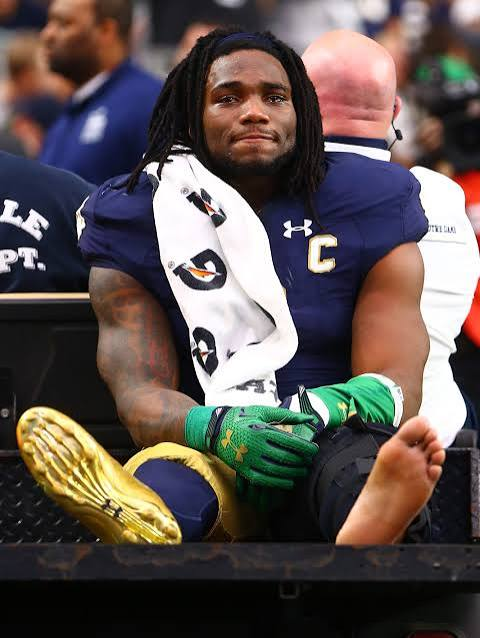
The most widely known public illustration of this injury is Notre Dame linebacker Jaylon Smith. According to public and sports-media reporting — this case has not been described in the peer-reviewed medical literature — a low block to the outside of his planted leg during the January 2016 Fiesta Bowl produced a hyperextension–varus injury with tearing of the ACL and LCL and an associated common peroneal nerve injury. A projected top-five NFL draft pick, he fell to the second round, was selected by the Dallas Cowboys, sat out his entire first professional season, and returned to competitive play in 2017 using a custom ankle-foot orthosis for persistent foot drop. He was later named to a Pro Bowl.
The ultimate extent of his nerve recovery is not verifiable from published sources; media accounts across subsequent seasons described varying degrees of returning function and changes in his bracing. The case is therefore most useful as an illustration of mechanism and time course — a lateral-sided, varus-hyperextension injury producing a nerve deficit that persisted well beyond a year and required orthotic management to permit high-level function — rather than as evidence about recovery odds. For prognosis, the cohort data below are the appropriate reference.
His case is not an outlier in sport. In a review of 448 peroneal nerve injuries, 84 (approximately 18%) were sports-related, with skiing (42 cases) and football (23 cases) the leading mechanisms; the typical pattern was a stretch/contusion injury rather than a clean transection.[3] Roughly one-third of all knee dislocations arise from sport.[4]
The anatomy: a nerve in the wrong place
The common peroneal (fibular) nerve is the smaller terminal division of the sciatic nerve. It travels along the medial border of the biceps femoris tendon, crosses the posterolateral knee, wraps around the fibular neck, and divides into superficial and deep branches. The deep branch supplies tibialis anterior, extensor hallucis longus, and extensor digitorum longus — the anterior compartment muscles responsible for dorsiflexion.[2]
Three anatomic features make this nerve uniquely vulnerable:
- Superficial, subcutaneous course at the fibular neck, where it lies directly against bone with minimal soft-tissue protection.[1][4]
- Tethering at the fibular neck, which converts a traction event at the knee into focal stretch across a fixed point, exposing the nerve to both compression and traction.[4]
- Fascicular architecture: compared with the tibial division, the peroneal division contains fewer, larger fascicles with relatively sparse protective connective tissue, making it less tolerant of stretch.[5]
The mechanism: varus and hyperextension
The nerve is most at risk during varus stress, particularly when the posterolateral corner (PLC) fails.[1] As the lateral side of the knee opens, the nerve — anchored distally at the fibula — is stretched over a lengthening distance.
MRI bone bruise mapping supports this. In a series of 108 MLKIs, 24 of 26 patients with CPN injury (92.3%) had anteromedial femoral condyle bone bruising, and all 20 patients with complete palsy had this finding. The pattern implies a hyperextension–varus mechanism from a high-energy blow to the anteromedial knee — exactly the geometry of a low block to a planted leg.[6] A grade 3 PLC injury carried the strongest association with nerve injury (OR 23.81), followed by anteromedial condyle bruising (OR 21.9) and documented dislocation (OR 3.45).[6]
Which injury patterns predict nerve injury
The lateral side of the knee is the critical variable:
- In 100 consecutive MLKIs, 31% of patients with a PLC injury had peroneal palsy versus 4% of those without a PLC injury.[7]
- The lateral collateral ligament was completely torn in 96% of PLC injuries. Midsubstance tears and fibular-sided avulsions — injuries closest to the nerve's course — were associated with higher rates of nerve injury than femoral-sided avulsions.[7]
- In a separate cohort, distal PLC injuries and biceps femoris avulsion were each significantly associated with clinical CPN palsy (each present in 9 of 11 palsy patients).[8]
- In a multicenter series of 221 MLKIs, the overall rate of clinical peroneal injury was 19.5%, and 100% of those patients had a PLC injury. Additionally, 95.3% had a complete ACL rupture.[9]
The practical message: an ACL plus PLC injury pattern — especially with a distal or fibular-sided lateral injury — is the signature combination behind post-traumatic foot drop.
Nerve injury severity determines the outcome
The single most important prognostic variable is whether the palsy is complete or partial at presentation.
- In a systematic review of 214 CPN palsies after traumatic knee dislocation, functional recovery (MRC ≥3/5) occurred in 38.4% after complete palsy, whereas 87.3% of partial palsies achieved full 5/5 recovery. Younger age predicted better neurologic recovery.[10]
- In the PLC pathoanatomy series, 100% of partial palsies regained function versus 12% of complete palsies.[7]
- In a Mayo cohort followed a mean 6.3 years, 38% with complete palsy versus 83% with partial palsy regained antigravity dorsiflexion.[11]
- In a registry of 247 knee dislocations, 43 patients (17%) had CPN paresis at admission; only 35% had improved at one year, and spontaneous improvement after 12 months was distinctly rare.[12]
- In a prospective multicenter series, the anatomic state of the nerve was the strongest prognostic factor; in roughly 25% of dislocations the nerve showed contusion–elongation over several centimeters, carrying a prognosis as poor as complete rupture.[13]
Chronic denervation compounds the problem: prolonged loss of dorsiflexion leads to equinus contracture and disuse atrophy, which further reduce the chance of meaningful reinnervation even if axons do regenerate.[2]
Treatment pathway
1. Neuropraxic or mild axonal injury — non-operative care with an ankle-foot orthosis and, critically, ongoing Achilles/gastrocnemius stretching to prevent fixed equinus.[4]
2. Neurolysis — often performed concurrently when the patient is already undergoing lateral ligamentous reconstruction; benefit in isolation is less clearly established. Good functional outcomes were reported in 85% after neurolysis for lower-intensity sports-related lesions.[4][3]
3. Nerve grafting or nerve transfer — considered for nerve discontinuity or significant axonal injury with non-resolving foot drop at approximately three months. Graft length drives prognosis: 70% good outcomes for grafts under 6 cm, 43% for 6–12 cm, and only 25% for 13–24 cm. Distal transfer of tibial motor fascicles to the deep peroneal nerve places healthy axons closer to the motor endplate for faster reinnervation.[4][3]
4. Posterior tibial tendon transfer — the most predictable salvage for persistent palsy. In 12 evaluable patients after knee dislocation, mean AOFAS score was 91 of 100, though dorsiflexion strength (118 Nm) remained well below the uninjured side (284 Nm). Transfer is recommended for foot drop persisting at least one year, and can be performed years later provided the donor muscle is healthy. The Bridle procedure — a modified posterior tibialis transfer with tri-tendon anastomosis — can return patients to brace-free ambulation and athletic function.[14][4][12]
A meta-analysis of individual participant data across 1284 patients found that traction/stretch mechanisms — the relevant category for MLKI — had their best functional results with tendon transfer compared with orthosis alone.[15]
Clinical bottom line
Foot drop after a multiligament knee injury is not incidental; it is the predictable consequence of a lateral-sided, varus-hyperextension injury pattern that stretches a tethered, poorly protected nerve across the fibular neck. Every MLKI warrants a documented dorsiflexion and sensory exam at presentation, with careful attention to preserved ankle inversion as the clinical discriminator favoring peroneal neuropathy over L5 radiculopathy.[5] When the palsy is complete, counsel realistically: fewer than half of these patients recover functional dorsiflexion, and the long-term plan should anticipate bracing, contracture prevention, and consideration of reconstructive or tendon transfer options.[10][11] The high-profile athlete cases underscore both realities — that even unlimited resources cannot guarantee nerve recovery, and that high-level function with an orthosis remains achievable.
SCHEDULE AN APPOINTMENT
Get an Expert Evaluation
If you have burning pain in the ball of your foot, numbness in your toes, or the feeling that you're always walking on a pebble — schedule an appointment with Dr. Sarang Desai for an expert evaluation and a clear treatment plan.
SCHEDULE AN EVALUATION
Dr. Sarang Desai
Orthopedic Surgeon – Foot & Ankle & Sports Medicine
Orthopedic Institute of North Texas
Phone: (972) 591-6468
Website: https://www.theachillesdoc.com
References
Nerve Injury Complicating Multiligament Knee Injury: Current Concepts and Treatment Algorithm. Mook WR, Ligh CA, Moorman CT, Leversedge FJ. The Journal of the American Academy of Orthopaedic Surgeons. 2013;21(6):343-54. doi:10.5435/JAAOS-21-06-343.
Distal Nerve Transfers for Foot Drop: A Systematic Review and Meta‐Analysis. Jerome JTJ, Surendran G, Kuppusamy T. Microsurgery. 2026;46(1):e70187. doi:10.1002/micr.70187.
Peroneal Nerve Injury Associated With Sports-Related Knee Injury. Cho D, Saetia K, Lee S, Kline DG, Kim DH. Neurosurgical Focus. 2011;31(5):E11. doi:10.3171/2011.9.FOCUS11187.
Common sports‐related nerve injuries seen by the electrodiagnostic medical consultant. Farag JI, McDougall AN, Catapano M. Muscle & Nerve. 2025;71(5):715-731. doi:10.1002/mus.28298.
Clinical Reasoning: A 51-Year-Old Woman With Acute Foot Drop. Rallis D, Skafida A, Alexopoulos G, et al. Neurology. 2015;84(7):e48-52. doi:10.1212/WNL.0000000000001261.
Examining the Bone Bruise Patterns in Multiligament Knee Injuries With Peroneal Nerve Injury. Moran J, Schneble CA, Katz LD, et al. The American Journal of Sports Medicine. 2022;50(6):1618-1626. doi:10.1177/03635465221087406.
The Pathoanatomy of Posterolateral Corner Ligamentous Disruption in Multiligament Knee Injuries Is Predictive of Peroneal Nerve Injury. Kahan JB, Li D, Schneble CA, et al. The American Journal of Sports Medicine. 2020;48(14):3541-3548. doi:10.1177/0363546520962503.
Distal Posterolateral Corner Injury in the Setting of Multiligament Knee Injury Increases Risk of Common Peroneal Palsy. Essilfie AA, Alaia EF, Bloom DA, et al. Knee Surgery, Sports Traumatology, Arthroscopy : Official Journal of the ESSKA. 2022;30(1):239-245. doi:10.1007/s00167-021-06469-z.
Relationship Between Peroneal Nerve and Anterior Cruciate Ligament Involvement in Multiligamentous Knee Injury: A Multicenter Study. Markus DH, Mojica ES, Bi A, et al. The Journal of the American Academy of Orthopaedic Surgeons. 2022;30(22):e1461-e1466. doi:10.5435/JAAOS-D-21-01252.
A Systematic Review of Peroneal Nerve Palsy and Recovery Following Traumatic Knee Dislocation. Woodmass JM, Romatowski NP, Esposito JG, Mohtadi NG, Longino PD. Knee Surgery, Sports Traumatology, Arthroscopy : Official Journal of the ESSKA. 2015;23(10):2992-3002. doi:10.1007/s00167-015-3676-7.
Is Peroneal Nerve Injury Associated With Worse Function After Knee Dislocation?. Krych AJ, Giuseffi SA, Kuzma SA, Stuart MJ, Levy BA. Clinical Orthopaedics and Related Research. 2014;472(9):2630-6. doi:10.1007/s11999-014-3542-9.
Posterior Tibial Tendon Transfer Improves Function for Foot Drop After Knee Dislocation. Molund M, Engebretsen L, Hvaal K, Hellesnes J, Ellingsen Husebye E. Clinical Orthopaedics and Related Research. 2014;472(9):2637-43. doi:10.1007/s11999-014-3533-x.
Common Peroneal Nerve Palsy Complicating Knee Dislocation and Bicruciate Ligaments Tears. Bonnevialle P, Dubrana F, Galau B, et al. Orthopaedics & Traumatology, Surgery & Research : OTSR. 2010;96(1):64-9. doi:10.1016/j.rcot.2009.12.004.
Current Concepts Review: Common Peroneal Nerve Palsy After Knee Dislocations. Dy CJ, Inclan PM, Matava MJ, Mackinnon SE, Johnson JE. Foot & Ankle International. 2021;42(5):658-668. doi:10.1177/1071100721995421.
Treatment Approach to Isolated Common Peroneal Nerve Palsy by Mechanism of Injury: Systematic Review and Meta-Analysis of Individual Participants' Data. Klifto KM, Azoury SC, Gurno CF, et al. Journal of Plastic, Reconstructive & Aesthetic Surgery : JPRAS. 2022;75(2):683-702. doi:10.1016/j.bjps.2021.09.040.


Comments