

Posterior Tibial Tendon Dysfunction (PTTD)
Updated: Aug 27
The Complete Guide to the Most Common Cause of Adult Flatfoot
DR. SARANG DESAI / COMPLETE GUIDE
---
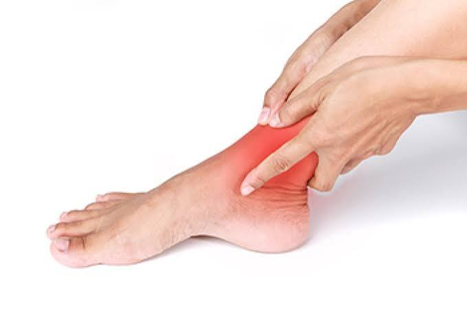
BY DR. SARANG DESAI | Fellowship-Trained Orthopedic Surgeon | Dallas–Fort Worth
If your arch is slowly flattening, your ankle aches on the inside, and your feet hurt in ways they never used to — you may have posterior tibial tendon dysfunction.
Posterior tibial tendon dysfunction (PTTD) is the most common cause of adult acquired flatfoot deformity. It happens when the main tendon supporting your arch becomes inflamed, degenerated, or torn — and gradually loses its ability to hold your foot up. Left untreated, the arch collapses, the heel drifts outward, and what started as a simple tendon problem becomes a complex three-dimensional deformity of the entire foot.
Here's the most important thing to know: PTTD is progressive. It does not stay the same. Caught early, it can usually be managed without surgery. Caught late, it may require major reconstruction or fusion. The difference between those two outcomes often comes down to how quickly the problem is recognized.
This is a complete guide to PTTD — what it is, why it happens, how to recognize it, how it's diagnosed, and every evidence-based treatment option available today.
---
PATIENT GUIDE
Key Takeaways
- PTTD is the most common cause of acquired flatfoot deformity in adults, and early recognition is the key to preventing long-term disability.
- The posterior tibial tendon is the main dynamic stabilizer of the arch — it absorbs shock at heel strike, stabilizes the foot at midstance, and generates force at push-off.
- Classic findings include pain on the inside of the ankle, a flattening arch, a "too many toes" sign, and difficulty performing a single-leg heel raise.
- Risk factors include obesity, high blood pressure, diabetes, corticosteroid exposure, collagen vascular disease, and excessive pronation. The condition is most common in women between ages 40 and 60.
- A structured nonoperative program of bracing plus high-repetition exercise achieved successful outcomes in 83% of patients with Stage I and II disease, with 89% satisfied — only 11% went on to surgery.
- Custom orthoses with individualized arch support outperform flat insoles for pain reduction in early-stage disease, according to a systematic review of randomized trials.
- Surgery is considered after 3 to 6 months of failed nonoperative treatment, and the procedure depends entirely on the stage.
- Expect at least a full year for maximum benefit after reconstruction. In one prospective study, outcomes improved at 6 months, continued improving through 1 year, and plateaued at 2 years.
- Most patients return to recreational activity after surgery — in one series, participation actually increased from 64% before surgery to 86% afterward.
---
PATIENT GUIDE
What Patients Need to Know First
Before diving into details, here are the practical realities that matter most:
This is not "just flat feet." Many people have flat feet their entire lives without problems. PTTD is different — it's a progressive collapse of an arch that used to be there. If your foot shape has changed in adulthood, that's a warning sign.
One foot is usually worse than the other. PTTD is typically asymmetric. If one foot looks flatter than the other, that asymmetry is meaningful.
The pain moves. Early on, it hurts on the inside of the ankle and arch. As the deformity worsens, it can start hurting on the outside of the ankle — because the heel bone tilts and pinches the tissues on the outer side. Patients often find this confusing. It's actually a sign of progression.
Stage matters more than anything else. The single most important question is: is the deformity flexible or fixed? A flexible deformity can be corrected with joint-preserving surgery. A fixed deformity usually requires fusion. This is why examination and imaging by a foot and ankle specialist is so important.
Waiting has real consequences. Untreated PTTD can progress to a painful, rigid flatfoot requiring extensive surgical reconstruction. Early management at Stage I can prevent the biomechanical changes that come with Stage II and beyond.
---
DEEP DIVE
Anatomy: What the Posterior Tibial Tendon Actually Does
The posterior tibial tendon starts from a muscle deep in your calf, travels down behind the inner ankle bone (the medial malleolus), curves around it like a rope around a pulley, and attaches to the navicular bone on the inside of your arch — with additional attachments to all three cuneiform bones and the second, third, and fourth metatarsals.
Its jobs are:
- Inverting the foot — turning the sole inward
- Plantarflexing the ankle — pointing the foot down
- Supporting the medial longitudinal arch — this is the big one
The tendon is the main dynamic stabilizing muscle of your arch. During walking, it performs three distinct functions:
1. Absorbs shock at heel strike
2. Stabilizes the foot during midstance
3. Generates force during heel lift and toe-off
Here's the critical point: the posterior tibial tendon has no true muscular backup. No other tendon can fully replace its function. When it fails, the arch has nothing holding it up dynamically — and it begins to collapse.
Why the tendon is vulnerable: There's a zone behind the inner ankle bone where the tendon's blood supply is relatively poor (a hypovascular zone). This area, combined with the mechanical stress of curving around the ankle bone, makes it a common site for degeneration and eventual tearing.
---
DEEP DIVE
The Cascade: How the Arch Collapses
Understanding this sequence explains why PTTD gets worse over time:
Step 1 — Tendon overload. Repetitive stress exceeds the tendon's capacity to repair itself. The tendon becomes inflamed and then degenerative.
Step 2 — Tendon elongation. As the tendon degenerates, it stretches. A stretched tendon can no longer hold the arch effectively.
Step 3 — Ligament failure. With the tendon no longer doing its job, the static supports of the arch — particularly the spring ligament and the deltoid ligament — take on loads they weren't designed for. Over time, they stretch and fail too.
Step 4 — Deformity. The arch flattens, the heel drifts into valgus (tilts outward), and the forefoot abducts (turns outward). This is the classic pes planovalgus deformity.
Step 5 — Rigidity. What began as a flexible deformity becomes fixed. Joint surfaces adapt to their abnormal positions. Arthritis develops.
Step 6 — Ankle involvement. In the most advanced stage, the deformity affects the ankle joint itself, causing the ankle to tilt into valgus.
This is why PTTD is now often called progressive collapsing foot deformity (PCFD) — the newer nomenclature better reflects that this is a whole-foot problem, not just a tendon problem.
---
DEEP DIVE
Biomechanics: What Research Shows About the Progression
A 2024 study in Gait & Posture used three-dimensional motion capture to compare walking mechanics between patients with Stage 1 PTTD, Stage 2 PTTD, and healthy controls. The findings were revealing:
- Stage 1 patients had minimal biomechanical differences compared to healthy controls
- Stage 2 patients showed dramatic changes — greater ankle plantarflexion and eversion, altered midfoot motion, and reduced midfoot abduction moments
- Critically, Stage 2 patients developed compensatory changes at the knee and hip — greater knee internal rotation, reduced knee external rotation moments, and altered hip mechanics
The authors' conclusion was direct and clinically important: PTTD is a progressive condition requiring early clinical management at Stage 1 to avoid the successive biomechanical changes associated with Stage 2.
Translation for patients: the window for simple treatment is early. Once the deformity develops, the problem is no longer confined to your foot — your knee and hip start compensating too.
---
PATIENT GUIDE
Causes and Risk Factors
PTTD develops from a combination of mechanical overload and biological vulnerability. Established risk factors include:
Body and metabolic factors:
- High body mass index (obesity) — increases load on the tendon with every step
- Diabetes mellitus — impairs tendon healing and blood supply
- Hypertension (high blood pressure) — associated with tendon degeneration
- Collagen vascular disease — including inflammatory arthritis conditions
Medication and exposure:
- Corticosteroid exposure — both injected and systemic steroids can weaken tendon tissue
Mechanical factors:
- Excessive pronation — a foot that rolls inward places continuous strain on the tendon
- Pre-existing flatfoot — a flexible flatfoot places the tendon at mechanical disadvantage
- Tight calf muscles (gastrocnemius contracture) — increases forces through the midfoot
- Overuse and overload — repetitive high-volume activity
Demographics:
The classic patient profile is a woman between 40 and 60 years old with obesity. That said, PTTD occurs in men, in younger patients, and in athletes — it just presents differently.
In athletes: PTTD tends to present earlier in the disease process, often as a Stage I overload problem without deformity. Runners, dancers, and court-sport athletes are most commonly affected. In these patients, the diagnosis is frequently mistaken for plantar fasciitis, a medial stress fracture, or a lingering ankle sprain.
---
PATIENT GUIDE
Symptoms: What PTTD Feels Like
Symptoms change as the condition progresses. This is one of the most useful diagnostic clues.
Early Symptoms (Stage I)
- Pain around the posteromedial ankle — behind and below the inner ankle bone
- Pain that radiates along the arch of the foot
- Swelling along the course of the tendon
- Pain worse with weight-bearing activity and better with rest
- No visible deformity — the foot still looks normal
- Difficulty or pain when trying to rise onto the toes of that foot alone
Progressive Symptoms (Stage II)
- All of the above, plus:
- Visible arch flattening — patients notice their foot "looks different"
- Heel tilting outward (hindfoot valgus)
- Shoes wearing unevenly or feeling too tight
- Inability to perform a single-leg heel raise
- Fatigue and aching after standing or walking
- The deformity is still flexible — it corrects when weight is off the foot
Advanced Symptoms (Stage III–IV)
- Rigid, fixed flatfoot — the deformity no longer corrects
- Pain on the outside of the ankle — from the heel bone pinching against the fibula (lateral impingement)
- Difficulty finding shoes that fit
- Pain even with limited walking
- In Stage IV, ankle pain and tilting from involvement of the ankle joint itself
The symptom shift to lateral pain is a red flag. When patients tell me their inner ankle pain has moved to the outer ankle, it usually means the deformity has progressed significantly.
---
DEEP DIVE
Staging: The Framework That Drives Treatment
The Johnson and Strom classification was the first system to categorize PTTD and remains the most widely used. It has been refined over time, and newer systems (progressive collapsing foot deformity classification) add detail — but the core concept is unchanged: stage determines treatment.
Stage | Tendon | Deformity | Key Exam Finding | Typical Treatment |
I | Inflamed/degenerative, normal length | None | Medial pain and swelling; painful single-leg heel raise | Nonoperative; debridement/tenosynovectomy if surgery needed |
II | Elongated or torn | Flexible flatfoot | Cannot perform single-leg heel raise; flexible hindfoot valgus | Nonoperative first; joint-sparing reconstruction (osteotomy + tendon transfer) |
III | Nonfunctional | Fixed hindfoot deformity, fixed forefoot supination | Rigid deformity; lateral impingement pain | Arthrodesis (subtalar, double, or triple fusion) |
IV | Nonfunctional | Fixed hindfoot plus ankle valgus | Ankle joint tilting on X-ray | Tibiocalcaneal or pantalar arthrodesis |
The critical clinical distinction is Stage II versus Stage III — flexible versus fixed. This single determination separates joint-preserving reconstruction from fusion surgery, and it has enormous implications for function and recovery.
---
PATIENT GUIDE
Diagnosis: How PTTD Is Confirmed
Physical Examination
A proper PTTD exam starts with the patient standing, which many providers skip. Key findings:
Standing inspection (from behind):
- Pes planovalgus deformity — flattened arch with heel tilted outward
- "Too many toes" sign — when viewing the foot from behind, more toes are visible on the affected side because the forefoot has drifted outward. This is one of the most reliable visual signs of PTTD.
- Heel valgus — the heel angles away from the midline
Single-limb heel raise test:
This is the single most important functional test. A healthy person should be able to perform five to ten single-leg heel raises without difficulty, and the heel should swing into inversion (turn inward) as they rise. In PTTD:
- The patient may be unable to perform the test at all
- It may be painful
- The heel fails to invert — a sign the posterior tibial tendon isn't functioning
Palpation:
- Swelling and tenderness along the tendon, particularly proximal to its attachment on the navicular tuberosity
Strength testing:
- Pain or weakness with resisted inversion of a plantarflexed foot — this isolates the posterior tibial tendon and separates it from the tibialis anterior
Flexibility assessment:
- Can the hindfoot deformity be manually corrected? This determines Stage II versus Stage III.
- Is there a tight calf (gastrocnemius contracture)? This is present in the majority of patients and must be addressed in any treatment plan.
Imaging
Weight-bearing X-rays are the essential first study. They must be standing — non-weight-bearing X-rays hide the deformity entirely. Key measurements include Meary's angle (arch alignment) and calcaneal pitch (heel bone angle). These are the same measurements used to assess surgical correction; in one series, Meary's angle improved from 11.5° to 7.0° and calcaneal pitch from 16.5° to 19.0° after reconstruction.
Weight-bearing CT is increasingly used at specialty centers because it shows the true three-dimensional deformity under load — something standard X-rays cannot capture.
MRI is the best study for evaluating the tendon itself. It shows tenosynovitis, tendon degeneration, partial tears, complete ruptures, and — importantly — the condition of the spring ligament, which is frequently injured alongside the tendon. MRI also assesses for cartilage damage and arthritis, which affects surgical planning.
Ultrasound is useful for dynamic assessment of the tendon and can be performed in the office. It's operator-dependent but valuable for confirming tenosynovitis and tears without radiation.
When each is most useful:
- X-ray: Always. Establishes alignment and stage. Screens for arthritis.
- MRI: When the diagnosis is unclear, when planning surgery, or when a tear is suspected.
- CT (weight-bearing preferred): For advanced deformity and complex surgical planning.
- Ultrasound: Quick office confirmation of tendon pathology; good for following treatment response.
---
DEEP DIVE
What Else Could It Be? Common Misdiagnoses
PTTD is frequently confused with other conditions. In my clinic, these are the most common mix-ups:
- Plantar fasciitis — Both cause arch pain. Plantar fasciitis pain is at the bottom of the heel and worst with the first steps in the morning. PTTD pain is behind the inner ankle bone and worsens with activity.
- Achilles tendinitis — Both cause posterior ankle pain and both can be worse in the morning. Achilles pain is in the back of the heel; PTTD pain is on the inner side.
- Tarsal tunnel syndrome — Nerve compression in the same region. Causes burning, tingling, or numbness rather than mechanical pain.
- Medial malleolar stress fracture — Bone pain, often in runners, with focal tenderness on the bone itself rather than the tendon.
- Deltoid ligament injury — Can coexist with PTTD, especially in advanced stages.
- Subtalar or midfoot arthritis — Can cause similar pain patterns and may coexist with PTTD.
- Flexor hallucis longus tendinopathy — Common in dancers, presents with posteromedial pain, but with great toe stiffness and crepitus.
Getting the diagnosis right matters because the treatments are entirely different. A patient treated for plantar fasciitis for a year while their arch quietly collapses has lost the window for simple intervention.
---
PATIENT GUIDE
Treatment: Nonoperative Management
The initial treatment for patients at any stage should be nonoperative. This is the consensus position, and the evidence supporting it is stronger than many patients realize.
The Landmark Nonoperative Study
The most important study on nonoperative PTTD treatment was published in Foot & Ankle International. Forty-seven consecutive patients with Stage I or II PTTD were treated with a structured protocol including:
- A short articulated ankle-foot orthosis or foot orthosis
- High-repetition exercises
- Aggressive plantarflexion activities
- An aggressive high-repetition home exercise program including calf stretching
The results were striking. After a median of only 10 physical therapy visits over 4 months:
- 83% of patients had successful subjective and functional outcomes
- 89% were satisfied
- Only 11% required surgery after nonoperative treatment failed
The success criteria were demanding — no more than 10% strength deficit, ability to perform 50 single-support heel rises with minimal pain, ability to walk 100 feet on the toes, and tolerance of 200 repetitions of home exercises per muscle group.
The take-home message: structured, aggressive, high-repetition rehabilitation works for the majority of patients with early-stage PTTD. But "structured and aggressive" is the key phrase. A generic three-exercise handout is not the same thing.
Activity Modification
Reducing provocative load is the foundation. This means:
- Temporarily reducing running mileage, court sports, and prolonged standing
- Substituting cycling, swimming, elliptical, and pool running to maintain fitness
- Avoiding barefoot walking on hard surfaces
- Avoiding unsupportive footwear (flip-flops, minimalist shoes, flat dress shoes)
Activity modification is not the same as complete rest. Tendons need load to heal — they just need the right load.
Orthotics and Bracing
This is the cornerstone of nonoperative treatment. Support for the medial longitudinal arch can be provided through:
- Custom foot orthoses with medial arch support and a medial heel post
- Taping — useful for short-term symptom control and as a trial before committing to orthoses
- A brace with an air cell to lift the arch
- Articulated ankle-foot orthosis (AFO) — for more significant symptoms or Stage II disease
- Arizona brace or gauntlet-style AFO — for advanced deformity or patients who are not surgical candidates
- Walking boot immobilization — reserved for severe, acute cases
The evidence on orthotics: A systematic review in Clinical Rehabilitation analyzed four randomized controlled trials (186 patients) and concluded that orthotic treatment may be effective in reducing pain in the early stages of PTTD. Two findings were particularly practical:
1. Foot orthoses combined with exercise programs worked better than orthoses alone
2. Foot orthoses with personalized internal longitudinal arch support were more effective than flat insoles or standard treatment for reducing pain
The authors also noted the limitations honestly — 75% of the included trials were at high risk of bias for blinding, and more research into individualized orthotics and high-intensity monitored exercise programs is needed. So the evidence supports orthoses, but it isn't definitive on optimal design.
Physical Therapy and Exercise
Physical therapy should focus on:
- Eccentric strengthening — eccentric exercises are recommended over concentric for posterior tibial tendinopathy
- High-repetition loading — the successful protocol used up to 200 repetitions per muscle group
- Calf stretching — gastrocsoleus contracture is nearly universal and must be addressed
- Posterior tibialis strengthening — resisted inversion with bands, progressing to weight-bearing
- Hip and core strengthening — because the compensations occur up the chain
- Balance and proprioception work
- Gait retraining
If you're interested in the principles behind tendon loading programs, the same concepts apply that I describe in my guide to the best exercises for tendon pain — progressive load, consistency, and patience.
Medications
- NSAIDs (ibuprofen, naproxen) — useful for the acute inflammatory phase. Reasonable short-term, not a long-term solution.
- Ice — helpful for acute symptoms
- Acetaminophen — for patients who cannot take NSAIDs
Corticosteroid Injections — A Word of Caution
Corticosteroid injections into or around the posterior tibial tendon remain controversial. The concern is significant: corticosteroid exposure is itself a known risk factor for posterior tibial tendon degeneration, and injecting steroid into a tendon that is already degenerating risks weakening it further and precipitating rupture.
My practice pattern — and this reflects expert clinical opinion rather than high-quality trial data — is to avoid intratendinous steroid injection in PTTD. If an injection is considered at all, it should be image-guided, placed in the tendon sheath rather than the tendon substance, and used sparingly. Patients who are offered steroid injections directly into this tendon should ask careful questions.
PRP and Regenerative Treatments
There is very limited high-quality evidence for platelet-rich plasma (PRP) in posterior tibial tendinopathy specifically. The large randomized trials of PRP in tendinopathy have focused on other tendons, and results there have been largely negative. Extrapolating from other tendons is not evidence.
Honest assessment: PRP for PTTD is not supported by high-quality data at this time. Patients considering it should understand they are paying out of pocket for a treatment without established benefit for this condition.
Weight Management
For patients with elevated BMI, weight reduction addresses one of the strongest modifiable risk factors. Every step transmits multiples of body weight through this tendon. This conversation is uncomfortable but it is clinically important, and I have seen patients meaningfully improve their symptoms through weight loss alone.
---
PATIENT GUIDE
Treatment: When Is Surgery Needed?
Surgical treatment is considered when three to six months of nonoperative treatment is ineffective. The specific procedure depends entirely on the stage.
Indications for Surgery
- Persistent pain despite 3 to 6 months of appropriate nonoperative treatment
- Progressive deformity despite bracing
- Functional limitation that materially affects work, activity, or quality of life
- Inability to tolerate bracing long-term
- Advanced deformity with lateral impingement pain
Surgery by Stage
Stage I — Debridement and Tenosynovectomy
The tendon sheath is opened, inflamed tissue is removed, and the tendon is debrided. In some cases, small tears are repaired. This is the least invasive surgical option. Some surgeons add a flexor digitorum longus (FDL) transfer or a medial calcaneal osteotomy if there is early hindfoot valgus, though this is individualized.
Stage II — Joint-Sparing Reconstruction
This is the most complex decision-making in PTTD surgery, and there is no consensus regarding a single surgical option for Stage II. Treatment involves combined soft tissue and bony procedures, typically selected from:
- Flexor digitorum longus (FDL) tendon transfer — the FDL tendon (which flexes the toes) is rerouted to substitute for the failed posterior tibial tendon
- Medial displacement calcaneal osteotomy (MDCO) — the heel bone is cut and shifted inward to correct hindfoot valgus
- Lateral column lengthening — corrects forefoot abduction
- Spring ligament repair or reconstruction — addresses the failed static support
- Gastrocnemius recession — lengthens the tight calf
- Cotton osteotomy (medial cuneiform) — corrects residual forefoot supination
- First TMT fusion — for instability of the medial column
The goal is to restore alignment while preserving joint motion. Soft tissue transfer alone does not correct the underlying deformity — this is a well-established principle. The bony correction is essential.
Stage III — Arthrodesis (Fusion)
When the deformity is fixed, the joints must be realigned and fused. Options include:
- Subtalar arthrodesis
- Double arthrodesis (subtalar and talonavicular)
- Triple arthrodesis (subtalar, talonavicular, and calcaneocuboid joints)
These are frequently combined with heel cord lengthening.
Stage IV — Extended Fusion
When the ankle joint is involved, tibiocalcaneal or pantalar arthrodesis is most commonly used. Some patients with preserved ankle cartilage may be candidates for deformity correction combined with ankle joint reconstruction, and total ankle replacement is being explored in selected cases. This is the most complex end of the spectrum. Patients who have developed ankle arthritis from long-standing deformity may want to review the 2026 AAOS ankle arthritis guidelines.
Surgical Outcomes
The outcome data for PTTD reconstruction are encouraging:
- A retrospective review of studies with 10-year follow-up in Stage II patients found 86% were totally satisfied or satisfied with minor reservations.
- A prospective Foot & Ankle International study of 24 patients undergoing FDL transfer, medial calcaneal osteotomy, lateral column lengthening, and gastrocnemius recession found all outcome measures showed statistically significant improvement, and patients were highly satisfied. Importantly, dynamometer testing showed no loss of plantarflexion strength despite the gastrocnemius recession — a common patient concern.
- A study on return to sport after medial displacement calcaneal osteotomy with FDL transfer found that athletic participation increased after surgery: 64.3% of patients participated in sports before surgery (averaging 1.4 hours/week) versus 85.7% after surgery (averaging 3.5 hours/week). Patients reported good scores for symptom tolerance (86.4%), pain tolerance (89.0%), daily living (96.1%), and sports performance (86.7%).
That last finding deserves emphasis: patients were more active after reconstruction than before. For patients worried that surgery means the end of their active life, this is important data.
Risks of Surgery
Every operation carries risk. For PTTD reconstruction, these include:
- Infection (superficial or deep)
- Nonunion or delayed union of osteotomies or fusions
- Nerve injury — particularly the sural nerve with lateral approaches
- Undercorrection or overcorrection of the deformity
- Hardware irritation — sometimes requiring removal
- Lateral column pain after lateral column lengthening
- Progression of arthritis in adjacent joints, particularly after fusion
- Recurrence of deformity
- Blood clots (DVT/PE)
- Prolonged swelling — often lasting many months
- Persistent stiffness — expected after fusion procedures
- Wound healing problems — higher risk in smokers and diabetics
Smoking is a major modifiable risk factor for nonunion and wound complications. Patients who smoke should stop before elective reconstruction.
---
PATIENT GUIDE
Recovery Timeline After PTTD Reconstruction
Recovery from PTTD surgery is longer than most patients expect. Here's a realistic framework. Note that specific protocols vary by surgeon and by which procedures are performed.
Weeks 0–2
- Splint or cast, strictly non-weight-bearing
- Elevation is critical — swelling control drives everything early
- Pain management, blood clot prevention
Weeks 2–6
- Transition to cast or boot, still non-weight-bearing in most protocols
- Sutures removed around 2 weeks
- Gentle toe and knee motion
- Upper body and contralateral leg conditioning
Weeks 6–10
- Progressive weight-bearing begins in a boot, as osteotomies and fusions heal
- X-rays confirm healing before advancing
- Begin formal physical therapy — range of motion, gentle strengthening, gait training
Weeks 10–16
- Transition out of the boot into supportive shoes with orthoses
- Progressive strengthening
- Stationary bike, pool work
- Most patients walking reasonably by 3 to 4 months
Months 4–6
- Full-time shoe wear with orthotic support
- Advanced strengthening and balance work
- Low-impact activity: walking, cycling, elliptical, swimming
- Return to sedentary or light-duty work is typically earlier — often 4 to 8 weeks depending on the job
Months 6–12
- Return to higher-impact activity as tolerated, individualized
- Running, if appropriate, generally not before 6 months and often later
- Continued strength gains
Months 12–24
- The prospective Foot & Ankle International data are clear on this: improvement is seen at 6 months, continues to improve at 1 year, and largely plateaus by 2 years. The authors concluded that a good functional outcome can be anticipated after the early postoperative period, but it should be expected to take at least one year for maximal benefit.
Set expectations accordingly. Patients who expect to feel great at three months are often discouraged. Patients who understand this is a one-year recovery do much better psychologically.
Swelling deserves special mention. Foot and ankle reconstruction produces swelling that can persist for 6 to 12 months, particularly at the end of the day. This is normal and not a sign of failure.
---
PATIENT GUIDE
Prevention: Protecting the Tendon
You cannot change your age or genetics, but several factors are modifiable:
- Maintain a healthy weight — the strongest modifiable risk factor
- Wear supportive footwear — replace running shoes every 300–500 miles; avoid prolonged barefoot walking on hard floors
- Use arch support if you overpronate — over-the-counter or custom, depending on symptoms
- Stretch your calves daily — a tight calf drives midfoot overload
- Strengthen the posterior tibialis — resisted inversion, single-leg heel raises, and balance work
- Progress training load gradually — the 10% rule for weekly mileage increases
- Control diabetes and blood pressure — both are associated with tendon degeneration
- Be cautious with corticosteroids — particularly injections near this tendon
- Don't ignore early symptoms — persistent inner ankle pain that lasts more than a few weeks deserves evaluation
---
PATIENT GUIDE
Frequently Asked Questions
Posterior Tibial Tendon Dysfunction: A Complete Guide
PATIENT GUIDE
Frequently Asked Questions
What is posterior tibial tendon dysfunction?
PTTD is a progressive condition in which the posterior tibial tendon — the main dynamic support for your arch — becomes inflamed, degenerated, elongated, or torn. Because the tendon can no longer hold the arch up, the foot gradually flattens, the heel tilts outward, and the forefoot drifts outward. It is the most common cause of adult acquired flatfoot deformity.
---
Is PTTD the same thing as flat feet?
No. Many people have flat feet their entire lives without pain or dysfunction. PTTD is an acquired, progressive collapse of an arch that used to be there. If your foot shape has changed in adulthood — especially on one side more than the other — that is not simply "flat feet."
---
Will PTTD go away on its own?
Untreated PTTD tends to progress rather than resolve. The tendon does not spontaneously regain its length or strength. However, symptoms can be well controlled in the majority of early-stage patients with a structured bracing and exercise program — one landmark protocol achieved successful outcomes in 83% of Stage I and II patients, with only 11% ultimately requiring surgery.
---
How do I know what stage I'm in?
Stage requires an examination and weight-bearing X-rays. The two questions that define it are: (1) Is there a visible deformity? If no, Stage I. (2) If there is a deformity, does it correct when the foot is manipulated or non-weight-bearing? If yes, Stage II. If it is rigid, Stage III. If the ankle joint is also tilted, Stage IV.
---
Can I still run with PTTD?
Early-stage patients often can, with modifications — supportive footwear, orthoses, reduced volume, softer surfaces, and a consistent strengthening program. What matters is whether running provokes escalating pain or progressive deformity. Running through worsening arch collapse is not advisable. This should be individualized with a foot and ankle specialist.
---
Do orthotics actually work for PTTD, or are they a waste of money?
They work, particularly early. A systematic review of four randomized trials found that orthotic treatment may reduce pain in early-stage PTTD, that orthoses plus exercise outperformed orthoses alone, and that custom orthoses with individualized arch support outperformed flat insoles. The evidence has limitations — most trials had high risk of bias for blinding — but the direction is consistent.
---
Should I get a cortisone shot for this?
Corticosteroid injection into or around the posterior tibial tendon is controversial. Corticosteroid exposure is itself a recognized risk factor for posterior tibial tendon degeneration, so injecting a degenerating tendon carries real risk of further weakening or rupture. If an injection is used at all, it should be image-guided, placed in the sheath rather than the tendon, and used sparingly.
---
Does PRP help posterior tibial tendinopathy?
There is very limited high-quality evidence for PRP in this specific tendon. Large randomized trials of PRP in tendinopathy have focused on other tendons, and results there have largely been negative. Extrapolation is not evidence. Patients considering PRP for PTTD should understand they would be paying out of pocket for a treatment without established benefit for this condition.
---
How long do I have to try nonoperative treatment before considering surgery?
Surgery is generally considered after 3 to 6 months of appropriate nonoperative treatment has failed. "Appropriate" is the operative word — a genuine trial means proper bracing or custom orthoses plus a structured, high-repetition strengthening program, not a few weeks in a drugstore insole.
---
What surgery will I need?
It depends entirely on stage. Stage I may need only tendon debridement. Stage II typically requires a combination of a tendon transfer (usually flexor digitorum longus) plus bony realignment — a medial displacement calcaneal osteotomy, sometimes lateral column lengthening, spring ligament repair, and gastrocnemius recession. Stage III requires fusion. Stage IV requires extended fusion involving the ankle. There is no single consensus procedure for Stage II — this is why surgeon experience matters.
---
Will a tendon transfer alone fix my flat foot?
No. This is one of the most important principles in PTTD surgery: soft tissue procedures alone do not correct the underlying bony deformity. Durable correction requires realigning the bones. A tendon transfer performed in isolation on a deformed foot is prone to stretching out and failing.
---
If my calf is lengthened during surgery, will I lose push-off strength?
This is a common and reasonable concern. In a prospective Foot & Ankle International study of patients undergoing FDL transfer, calcaneal osteotomy, lateral column lengthening, and gastrocnemius recession, dynamometer testing showed no loss of plantarflexion strength. All outcome measures improved significantly and patients were highly satisfied.
---
How long is recovery after PTTD surgery?
Plan on a year. Most protocols involve non-weight-bearing for roughly 6 weeks, progressive weight-bearing in a boot through about 10 weeks, and transition to shoes with orthoses around 3 to 4 months. Prospective data show improvement at 6 months, continued improvement through 1 year, and a plateau by 2 years — maximum benefit should be expected to take at least one year.
---
Can I go back to sports after PTTD reconstruction?
Most patients can, and the data are more encouraging than many expect. In a study of patients undergoing medial displacement calcaneal osteotomy with FDL transfer, sports participation increased from 64.3% before surgery (1.4 hours/week) to 85.7% after surgery (3.5 hours/week), with high scores for pain tolerance and sports performance.
---
What happens if I just live with it?
Some patients do, particularly older or lower-demand patients, and a well-fitted gauntlet-style brace (such as an Arizona brace) can be an effective long-term strategy. The tradeoff is that deformity tends to progress, flexible deformity becomes rigid, and the surgical options narrow from joint-preserving reconstruction to fusion. A 2024 three-dimensional gait analysis showed that by Stage II, compensatory changes had already developed at the knee and hip — the problem stops being confined to the foot.
---
Why does my pain feel like it moved to the outside of my ankle?
This is a sign of progression. As the heel tilts outward, the heel bone begins impinging against the fibula and the soft tissues on the outer side of the ankle — called lateral impingement. Patients often interpret this as a new, unrelated problem. It is actually the same problem, further along.
---
---
PATIENT GUIDE
Myth vs. Fact
---
MYTH — Flat feet always cause pain, and painful flat feet are always PTTD.
FACT — Plenty of people have lifelong flat feet with no symptoms whatsoever. PTTD is specifically an acquired, progressive collapse driven by failure of the posterior tibial tendon. Distinguishing congenital flexible flatfoot from acquired deformity changes the entire treatment plan, and a systematic review has specifically highlighted that "adult acquired flatfoot deformity" and "posterior tibial tendon dysfunction" are not perfectly interchangeable terms even in the research literature.
---
MYTH — If it hurts on the inside of my ankle, it's probably an old ankle sprain.
FACT — Persistent posteromedial ankle pain with swelling along the tendon, pain on resisted inversion, and difficulty with a single-leg heel raise points to the posterior tibial tendon — not the lateral ligaments injured in a typical sprain. Attributing PTTD to a lingering sprain is one of the more common reasons diagnosis gets delayed.
---
MYTH — Non-weight-bearing X-rays are good enough to evaluate a collapsing arch.
FACT — Non-weight-bearing X-rays can hide the deformity almost entirely. Alignment must be assessed under load. Weight-bearing X-rays are essential, and weight-bearing CT is increasingly used at specialty centers to capture the true three-dimensional deformity.
---
MYTH — A cortisone shot is a safe, easy first step.
FACT — Corticosteroid exposure is a recognized risk factor for posterior tibial tendon degeneration. Injecting steroid into an already-degenerating, weight-bearing tendon risks further weakening and rupture. This is a situation where the easy intervention carries real downside.
---
MYTH — Rest is the treatment. Stay off it and it will settle down.
FACT — Complete rest deconditions the tendon without restoring its capacity. The best-documented nonoperative protocol was built on the opposite principle: aggressive, high-repetition loading — up to 200 repetitions per muscle group at home, plantarflexion work, and calf stretching — alongside bracing. That program produced 83% success and 89% satisfaction in Stage I and II disease.
---
MYTH — Surgery for flatfoot means having your foot fused.
FACT — Fusion is the answer for fixed (Stage III–IV) deformity. For flexible Stage II deformity, the goal is the opposite: joint-preserving realignment with osteotomies, tendon transfer, and ligament repair, which maintains motion. This is precisely why the flexible-versus-rigid determination is the single most consequential part of the examination.
---
MYTH — Surgery will end my athletic life.
FACT — In one series of patients undergoing medial displacement calcaneal osteotomy with FDL transfer, athletic participation rose from 64.3% preoperatively to 85.7% postoperatively, and weekly activity time more than doubled. Long-term data in Stage II patients at 10 years showed 86% were satisfied or satisfied with minor reservations.
---
MYTH — If I'm not better by three months after surgery, something went wrong.
FACT — Prospective data show that outcomes improve at 6 months, keep improving through 1 year, and plateau around 2 years. Persistent swelling for 6 to 12 months is normal. At least a year should be expected for maximal benefit — this is a slow, forward-moving recovery, not a fast one.
---
MYTH — Waiting won't hurt anything. I can always have surgery later.
FACT — Waiting changes what surgery is possible. Flexible deformities become rigid, cartilage wears, and joint-sparing reconstruction gives way to fusion. Three-dimensional gait analysis has shown that Stage II patients have already developed compensatory changes at the knee and hip, whereas Stage I patients walk nearly like healthy controls — which is exactly why early management is emphasized.
---
---
EVIDENCE REVIEW
Research Summary
---
STRUCTURED NONOPERATIVE TREATMENT WORKS FOR MOST EARLY-STAGE PATIENTS.
In a Foot & Ankle International study of 47 consecutive patients with Stage I or II PTTD treated with an orthosis plus an aggressive high-repetition exercise program, 83% had successful subjective and functional outcomes and 89% were satisfied after a median of only 10 therapy visits over 4 months. Only 11% went on to surgery. The success bar was high — 50 single-support heel rises with minimal pain and no more than a 10% strength deficit. Plain-English takeaway: the program has to be aggressive and consistent, but when it is, most patients avoid the operating room.
---
CUSTOM ARCH SUPPORT PLUS EXERCISE BEATS EITHER ALONE.
A Clinical Rehabilitation systematic review of four randomized trials (186 patients) concluded that orthotic treatment may reduce pain in early-stage PTTD, that orthoses combined with exercise outperformed orthoses alone, and that individualized longitudinal arch support outperformed flat insoles or standard care. The authors were candid that 75% of trials carried high risk of bias for blinding, so this is supportive rather than definitive evidence.
---
THE DEFORMITY IS NOT JUST A FOOT PROBLEM BY STAGE II.
A 2024 three-dimensional gait analysis found that Stage 1 patients differed minimally from healthy controls, while Stage 2 patients showed markedly altered ankle and midfoot mechanics plus compensatory changes at the knee and hip. The authors specifically called for early clinical management at Stage 1 to prevent the successive changes associated with Stage 2. This is the strongest mechanistic argument for not waiting.
---
STAGE II SURGERY REQUIRES BOTH SOFT TISSUE AND BONE WORK — AND THERE IS NO SINGLE AGREED-UPON OPERATION.
The AAOS review of adult acquired flatfoot deformity is explicit that no consensus exists regarding a single surgical option for Stage II, and that management combines soft tissue and bony procedures. Soft tissue transfer alone does not correct the deformity. Practically, this means the specific combination should be tailored to the individual foot by a surgeon who does these regularly.
---
RECONSTRUCTION IMPROVES FUNCTION WITHOUT SACRIFICING PUSH-OFF STRENGTH.
A prospective Foot & Ankle International study of 24 patients undergoing FDL transfer, medial calcaneal osteotomy, lateral column lengthening, and gastrocnemius recession found statistically significant improvement in all outcome measures, high satisfaction, radiographic correction (Meary's angle 11.5° → 7.0°; calcaneal pitch 16.5° → 19.0°), and no measurable loss of plantarflexion strength on dynamometry.
---
RECOVERY IS MEASURED IN MONTHS TO A YEAR — NOT WEEKS.
The same prospective data showed improvement at 6 months, continued improvement at 1 year, and a plateau by 2 years, with the explicit conclusion that at least one year should be expected for maximal benefit. Long-term Stage II follow-up at 10 years showed 86% satisfied or satisfied with minor reservations.
---
ATHLETIC PARTICIPATION OFTEN INCREASES AFTER SURGERY.
After medial displacement calcaneal osteotomy with FDL transfer, sports participation rose from 64.3% preoperatively (1.4 hours/week) to 85.7% postoperatively (3.5 hours/week), with patient-reported scores of 86.4% for symptom tolerance, 89.0% for pain tolerance, 96.1% for daily living, and 86.7% for sports performance.
---
---
PATIENT GUIDE
Conclusion
Posterior tibial tendon dysfunction is one of the few foot and ankle conditions where the timing of diagnosis genuinely changes the treatment you're eligible for.
The core messages:
- PTTD is the most common cause of adult acquired flatfoot deformity, and it is progressive.
- The single most important clinical question is whether the deformity is flexible or fixed — that determines whether you're a candidate for joint-preserving reconstruction or fusion.
- Nonoperative treatment should be tried first at every stage, and when it's done properly — real arch support plus aggressive, high-repetition strengthening — the majority of early-stage patients succeed without surgery.
- Custom orthoses combined with exercise have the best supporting evidence among nonoperative options.
- Corticosteroid injections into this tendon warrant real caution, and PRP is not supported by high-quality evidence for this condition.
- When surgery is needed, outcomes are good — with high long-term satisfaction, preserved push-off strength, and often increased athletic participation — but recovery takes a full year.
If your arch is flattening, if one foot looks different from the other, if the inside of your ankle aches with activity, or if you can no longer rise onto the toes of one foot — get evaluated. Persistent posteromedial ankle pain lasting more than a few weeks is not something to wait out. This condition rewards early attention more than almost any other in foot and ankle surgery.
---
---
ABOUT THE AUTHOR
Dr. Sarang Desai
Dr. Sarang Desai is a fellowship-trained orthopedic surgeon specializing in foot and ankle surgery and sports medicine, based in the Dallas–Fort Worth area. A former All-American athlete at the University of Texas, Dr. Desai brings a unique understanding of what it takes to recover from injury and return to peak performance.
Dr. Desai has served as a professional sports team physician and currently owns multiple professional sports teams, giving him a distinctive perspective on athlete care and return-to-play decisions that few surgeons can offer. He is the inventor of multiple orthopedic implants and surgical devices and has authored numerous peer-reviewed scientific publications.
With more than 15 years of clinical experience, Dr. Desai treats everyone from weekend warriors and recreational athletes to collegiate and professional competitors. His practice is built on evidence-based, personalized care — combining the latest research with real-world clinical expertise to help every patient achieve the best possible outcome.
---
---
SCHEDULE AN APPOINTMENT
Get an Expert Evaluation
If you have persistent pain on the inside of your ankle, notice your arch flattening, have been told you have "flat feet" that are getting worse, or would like an expert opinion on whether you need surgery — schedule an appointment with Dr. Sarang Desai.
SCHEDULE AN EVALUATION
Dr. Sarang Desai
Orthopedic Surgeon – Foot & Ankle & Sports Medicine
Orthopedic Institute of North Texas
Schedule an appointment: (972) 591-6468 Locations: McKinney, TX | Flower Mound, TX
Website: https://www.theachillesdoc.com
Locations:
- McKinney, Texas
- Flower Mound, Texas
Call our office or request an appointment online to be seen promptly.
---
RELATED READING
Related Articles
- Plantar Fasciitis: Causes, Symptoms, Treatment, and Recovery — the condition most often confused with early PTTD
- Achilles Tendinitis: Causes, Symptoms, Treatment, and Recovery — the other major tendinopathy around the ankle
- Peroneal Tendon Injuries — the tendons on the opposite side of the ankle
- Ankle Arthritis Treatment: What the 2026 AAOS Guidelines Mean for You — relevant for advanced Stage IV disease
- Best Exercises for Tendon Pain — the loading principles that apply to PTTD rehab
---
ARTICLE INFORMATION
Medical Disclaimer
This article is intended for educational purposes only and should not replace an evaluation by a qualified healthcare professional. Every injury is unique, and treatment recommendations should be individualized based on your symptoms, examination, imaging findings, and goals.


Comments